MRI Features of Hepatocellular Carcinoma Related to Biologic Behavior
- Affiliations
-
- 1Department of Radiology, Gangnam Severance Hospital, Yonsei University College of Medicine, Seoul 135-720, Korea.
- 2Department of Radiology, Severance Hospital, Yonsei University College of Medicine, Seoul 120-752, Korea. gafield2@yuhs.ac
- KMID: 2155519
- DOI: http://doi.org/10.3348/kjr.2015.16.3.449
Abstract
- Imaging studies including magnetic resonance imaging (MRI) play a crucial role in the diagnosis and staging of hepatocellular carcinoma (HCC). Several recent studies reveal a large number of MRI features related to the prognosis of HCC. In this review, we discuss various MRI features of HCC and their implications for the diagnosis and prognosis as imaging biomarkers. As a whole, the favorable MRI findings of HCC are small size, encapsulation, intralesional fat, high apparent diffusion coefficient (ADC) value, and smooth margins or hyperintensity on the hepatobiliary phase of gadoxetic acid-enhanced MRI. Unfavorable findings include large size, multifocality, low ADC value, non-smooth margins or hypointensity on hepatobiliary phase images. MRI findings are potential imaging biomarkers in patients with HCC.
MeSH Terms
-
Aged
Biological Products
Biomarkers, Tumor
Carcinoma, Hepatocellular/diagnosis/pathology/*radiography
Contrast Media
Gadolinium DTPA
Humans
Liver Neoplasms/diagnosis/pathology/*radiography
Magnetic Resonance Imaging/*methods
Male
Middle Aged
Neoplasm Staging/methods
Prognosis
Biological Products
Biomarkers, Tumor
Contrast Media
Gadolinium DTPA
Figure
-
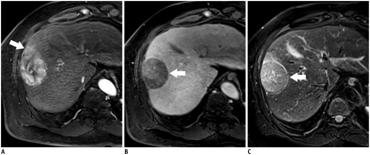
Fig. 1 55-year-old man with encapsulated progressed HCC.A. T1-weighted three-dimensional gradient echo image with fat suppression (TR/TE/FA = 2.5 ms/1 ms/11°) in late hepatic arterial phase after administration of gadoxetic acid shows hyperenhancing mass with hyperemia of surrounding liver parenchyma (arrow) in segment 8. B. Mass is hypointense on transitional phase with thin capsule appearance (arrow). Note that relatively high enhancement of background parenchyma on transitional phase may obscure capsular enhancement and reduce confidence of reader. C. Fat-suppressed fast spin echo T2-weighted image shows slight hyperintensity and hypointense capsule (arrow). FA = flip angle, HCC = hepatocellular carcinoma, TE = echo time, TR = repetition time
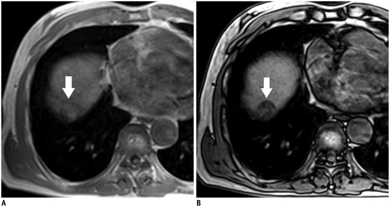
Fig. 2 76-year-old man with fat-containing HCC.A, B. Axial dual-echo gradient echo images (TR/TE = 4/1.2 ms, in-phase; 2.4 ms, opposed-phase) show mass in dome of liver. Signal loss (arrow) of mass on opposed-phase (B) compared to in-phase (A) indicates intralesional fat. Presence of intralesional fat permits confident diagnosis of HCC. HCC = hepatocellular carcinoma, TE = echo time, TR = repetition time
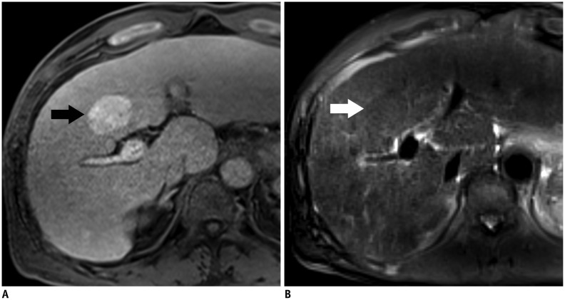
Fig. 3 71-year-old man with HCC showing T1 hyperintensity.A. Unenhanced fat-saturated T1-weighted three-dimensional gradient echo image with fat suppression (TR/TE/FA = 2.5 ms/0.9 ms/11°) shows hyperintense mass (arrow) in segment 4. B. Mass is barely visible on fat-suppressed fast spin echo T2-weighted imaging due to isointense signal (arrow). FA = flip angle, HCC = hepatocellular carcinoma, TE = echo time, TR = repetition time
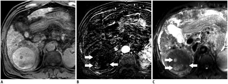
Fig. 4 56-year-old man with HCC showing nodule-in-nodule appearance.A. Pre-contrast T1-weighted three-dimensional GRE image shows hypointense inner nodules within hyperintense outer nodule, consistent with nodule-in-nodule architecture. B. Subtracted arterial phase image shows hyper-enhancement of inner nodules (arrows). C. Inner nodules (arrows) exhibit hyperintensity relative to outer nodule and surrounding liver on fat-suppressed fast spin echo T2-weighted image. Outer nodule is hypointense. GRE = gradient echo, HCC = hepatocellular carcinoma
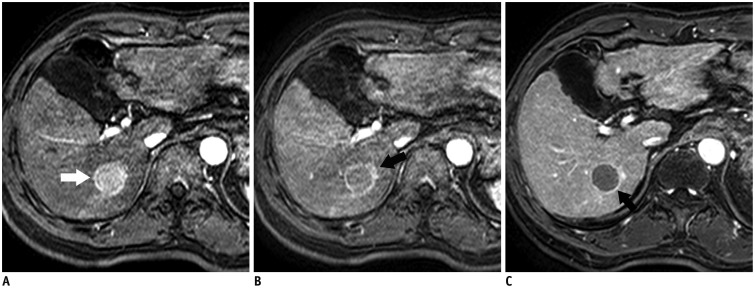
Fig. 5 66-year-old man with HCC showing corona enhancement.A, B. T1-weighted three-dimensional GRE image with fat suppression (TR/TE/FA = 4.5 ms/2.1 ms/15°) in (A) early and (B) late hepatic arterial phase after administration of gadolinium-based contrast agent shows hyper-enhancing mass (arrow) in segment 6. Notice irregular circumferential enhancement (arrow) in liver parenchyma around mass in late hepatic arterial phase. C. Enhancement of perilesional parenchyma fades in portal venous phase. Transient enhancement of perilesional parenchyma is known as corona enhancement. Note capsular appearance of mass (arrow). FA = flip angle, GRE = gradient echo, HCC = hepatocellular carcinoma, TE = echo time, TR = repetition time
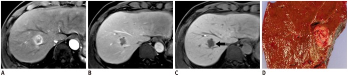
Fig. 6 51-year-old man with HCC and microvascular invasion.A. Gadoxetic acid-enhanced T1-weighted three-dimensional GRE sequence (TR/TE/FA = 3.4 ms/1.7 ms/15°) acquired in late hepatic arterial phase shows heterogeneous mass at segment 8 of liver. B. Transitional phase image at three minutes depicts hypointense mass. C. Margin of mass is non-smooth or lobulated on hepatobiliary phase imaging acquired 20 minutes after injection (arrow). D. Gross pathology photograph of resected specimen reveals confluent, multinodular type HCC. Histopathologic examination proves frequent microvascular invasion. FA = flip angle, GRE = gradient echo, HCC = hepatocellular carcinoma, TE = echo time, TR = repetition time
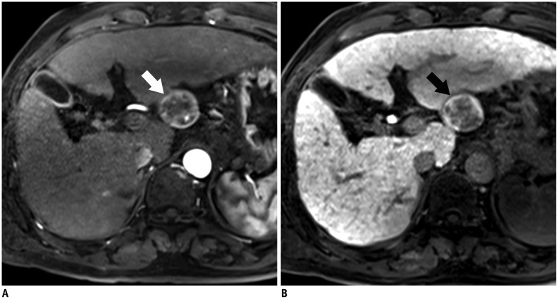
Fig. 7 64-year-old man with HCC showing hyperintensity in hepatobiliary phase.A. Gadoxetic acid-enhanced T1-weighted three-dimensional GRE image (TR/TE/FA = 3.4 ms/1.7 ms/15°) in late arterial phase shows exophytic, hyperenhancing mass (arrow) in left posterior liver. B. In hepatobiliary phase, mass is hyperintense with central hypointense areas (arrow). FA = flip angle, GRE = gradient echo, HCC = hepatocellular carcinoma, TE = echo time, TR = repetition time
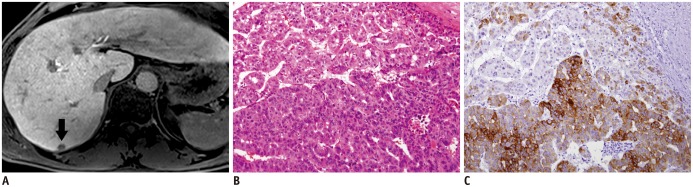
Fig. 8 51-year-old man with HCC.A. Hepatobiliary phase image acquired 20 minutes after injection shows hypointense nodule in segment 7 of liver (arrow). B. Nuclear grade III trabecular and pseudoglandular HCC with hepatic cells was confirmed (hematoxylin and eosin staining, × 100). C. Immunoreactivity of keratin 19 was strongly positive (× 100). Cell density ratio was 2.1. HCC = hepatocellular carcinoma
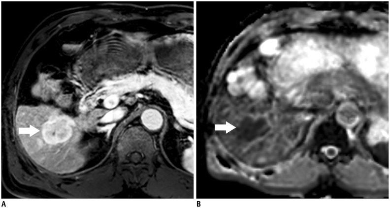
Fig. 9 63-year-old man with HCC showing restricted diffusion.A. Gadoxetic acid-enhanced T1-weighted three-dimensional GRE sequence (TR/TE/FA = 3.4 ms/1.7 ms/15°) in late arterial phase shows hyperenhancing mass (arrow) in right lobe of liver. B. Apparent diffusion coefficient map shows hypointensity (arrow) suggesting restricted diffusion. Restricted diffusion is highly suggestive of malignancy, but is not specific for HCC. FA = flip angle, GRE = gradient echo, HCC = hepatocellular carcinoma, TE = echo time, TR = repetition time
Cited by 1 articles
-
Value of Image Subtraction for the Identification of Hepatocellular Carcinoma Capsule on Gadoxetic Acid-Enhanced MRI
Hyunjung Kim, Jhii-Hyun Ahn, Jin Sil Moon, Seung-Whan Cha
J Korean Soc Radiol. 2018;79(6):340-347. doi: 10.3348/jksr.2018.79.6.340.
Reference
-
1. Pons F, Varela M, Llovet JM. Staging systems in hepatocellular carcinoma. HPB (Oxford). 2005; 7:35–41. PMID: 18333159.
Article2. Maida M, Orlando E, Cammà C, Cabibbo G. Staging systems of hepatocellular carcinoma: a review of literature. World J Gastroenterol. 2014; 20:4141–4150. PMID: 24764652.
Article3. Bruix J, Llovet JM. Prognostic prediction and treatment strategy in hepatocellular carcinoma. Hepatology. 2002; 35:519–524. PMID: 11870363.
Article4. Llovet JM. Updated treatment approach to hepatocellular carcinoma. J Gastroenterol. 2005; 40:225–235. PMID: 15830281.
Article5. Biomarkers Definitions Working Group. Biomarkers and surrogate endpoints: preferred definitions and conceptual framework. Clin Pharmacol Ther. 2001; 69:89–95. PMID: 11240971.6. Ludwig JA, Weinstein JN. Biomarkers in cancer staging, prognosis and treatment selection. Nat Rev Cancer. 2005; 5:845–856. PMID: 16239904.
Article7. Wagner JA, Williams SA, Webster CJ. Biomarkers and surrogate end points for fit-for-purpose development and regulatory evaluation of new drugs. Clin Pharmacol Ther. 2007; 81:104–107. PMID: 17186007.
Article8. Mishra A, Verma M. Cancer biomarkers: are we ready for the prime time? Cancers (Basel). 2010; 2:190–208. PMID: 24281040.
Article9. Martins A, Cortez-Pinto H, Marques-Vidal P, Mendes N, Silva S, Fatela N, et al. Treatment and prognostic factors in patients with hepatocellular carcinoma. Liver Int. 2006; 26:680–687. PMID: 16842324.
Article10. Chen WT, Chau GY, Lui WY, Tsay SH, King KL, Loong CC, et al. Recurrent hepatocellular carcinoma after hepatic resection: prognostic factors and long-term outcome. Eur J Surg Oncol. 2004; 30:414–420. PMID: 15063895.
Article11. Khatri G, Merrick L, Miller FH. MR imaging of hepatocellular carcinoma. Magn Reson Imaging Clin N Am. 2010; 18:421–450. xPMID: 21094448.
Article12. Choi JY, Lee JM, Sirlin CB. CT and MR imaging diagnosis and staging of hepatocellular carcinoma: part I. Development, growth, and spread: key pathologic and imaging aspects. Radiology. 2014; 272:635–654. PMID: 25153274.
Article13. The Cancer of the Liver Italian Program (CLIP) Investigators. Prospective validation of the CLIP score: a new prognostic system for patients with cirrhosis and hepatocellular carcinoma. Hepatology. 2000; 31:840–845. PMID: 10733537.14. Pomfret EA, Washburn K, Wald C, Nalesnik MA, Douglas D, Russo M, et al. Report of a national conference on liver allocation in patients with hepatocellular carcinoma in the United States. Liver Transpl. 2010; 16:262–278. PMID: 20209641.
Article15. Giuliante F, Ardito F, Pinna AD, Sarno G, Giulini SM, Ercolani G, et al. Liver resection for hepatocellular carcinoma ≤3 cm: results of an Italian multicenter study on 588 patients. J Am Coll Surg. 2012; 215:244–254. PMID: 22634119.
Article16. Gouw AS, Balabaud C, Kusano H, Todo S, Ichida T, Kojiro M. Markers for microvascular invasion in hepatocellular carcinoma: where do we stand? Liver Transpl. 2011; 17(Suppl 2):S72–S80. PMID: 21714066.
Article17. Kim KA, Kim MJ, Jeon HM, Kim KS, Choi JS, Ahn SH, et al. Prediction of microvascular invasion of hepatocellular carcinoma: usefulness of peritumoral hypointensity seen on gadoxetate disodium-enhanced hepatobiliary phase images. J Magn Reson Imaging. 2012; 35:629–634. PMID: 22069244.
Article18. Chandarana H, Robinson E, Hajdu CH, Drozhinin L, Babb JS, Taouli B. Microvascular invasion in hepatocellular carcinoma: is it predictable with pretransplant MRI? AJR Am J Roentgenol. 2011; 196:1083–1089. PMID: 21512074.
Article19. Efremidis SC, Hytiroglou P, Matsui O. Enhancement patterns and signal-intensity characteristics of small hepatocellular carcinoma in cirrhosis: pathologic basis and diagnostic challenges. Eur Radiol. 2007; 17:2969–2982. PMID: 17618439.
Article20. Roskams T, Kojiro M. Pathology of early hepatocellular carcinoma: conventional and molecular diagnosis. Semin Liver Dis. 2010; 30:17–25. PMID: 20175030.
Article21. Nakashima Y, Nakashima O, Hsia CC, Kojiro M, Tabor E. Vascularization of small hepatocellular carcinomas: correlation with differentiation. Liver. 1999; 19:12–18. PMID: 9928760.
Article22. Kondo F, Kondo Y, Nagato Y, Tomizawa M, Wada K. Interstitial tumour cell invasion in small hepatocellular carcinoma. Evaluation in microscopic and low magnification views. J Gastroenterol Hepatol. 1994; 9:604–612. PMID: 7865720.
Article23. Kojiro M. Histopathology of liver cancers. Best Pract Res Clin Gastroenterol. 2005; 19:39–62. PMID: 15757804.
Article24. Roncalli M, Park YN, Di Tommaso L. Histopathological classification of hepatocellular carcinoma. Dig Liver Dis. 2010; 42(Suppl 3):S228–S234. PMID: 20547308.
Article25. International Consensus Group for Hepatocellular Neoplasia. Pathologic diagnosis of early hepatocellular carcinoma: a report of the international consensus group for hepatocellular neoplasia. Hepatology. 2009; 49:658–664. PMID: 19177576.26. Park YN. Update on precursor and early lesions of hepatocellular carcinomas. Arch Pathol Lab Med. 2011; 135:704–715. PMID: 21631263.
Article27. Choi YS, Rhee H, Choi JY, Chung YE, Park YN, Kim KW, et al. Histological characteristics of small hepatocellular carcinomas showing atypical enhancement patterns on gadoxetic acid-enhanced MR imaging. J Magn Reson Imaging. 2013; 37:1384–1391. PMID: 23172629.
Article28. Sano K, Ichikawa T, Motosugi U, Sou H, Muhi AM, Matsuda M, et al. Imaging study of early hepatocellular carcinoma: usefulness of gadoxetic acid-enhanced MR imaging. Radiology. 2011; 261:834–844. PMID: 21998047.
Article29. Choi JY, Lee JM, Sirlin CB. CT and MR imaging diagnosis and staging of hepatocellular carcinoma: part II. Extracellular agents, hepatobiliary agents, and ancillary imaging features. Radiology. 2014; 273:30–50. PMID: 25247563.
Article30. Kogita S, Imai Y, Okada M, Kim T, Onishi H, Takamura M, et al. Gd-EOB-DTPA-enhanced magnetic resonance images of hepatocellular carcinoma: correlation with histological grading and portal blood flow. Eur Radiol. 2010; 20:2405–2413. PMID: 20490505.
Article31. Bartolozzi C, Battaglia V, Bargellini I, Bozzi E, Campani D, Pollina LE, et al. Contrast-enhanced magnetic resonance imaging of 102 nodules in cirrhosis: correlation with histological findings on explanted livers. Abdom Imaging. 2013; 38:290–296. PMID: 23053453.
Article32. Kim MJ, Lee M, Choi JY, Park YN. Imaging features of small hepatocellular carcinomas with microvascular invasion on gadoxetic acid-enhanced MR imaging. Eur J Radiol. 2012; 81:2507–2512. PMID: 22137613.
Article33. Ooka Y, Kanai F, Okabe S, Ueda T, Shimofusa R, Ogasawara S, et al. Gadoxetic acid-enhanced MRI compared with CT during angiography in the diagnosis of hepatocellular carcinoma. Magn Reson Imaging. 2013; 31:748–754. PMID: 23218794.
Article34. Okusaka T, Okada S, Ueno H, Ikeda M, Shimada K, Yamamoto J, et al. Satellite lesions in patients with small hepatocellular carcinoma with reference to clinicopathologic features. Cancer. 2002; 95:1931–1937. PMID: 12404287.
Article35. Theise ND, Curado MP, Franceschi S, Hytiroglou P, Kudo M, Park YN, et al. Hepatocellular carcinoma. In : Bosman FT, Carneiro F, Hruban RH, Theise ND, editors. WHO Classification of Tumors of the Digestive System. 4th ed. Lyon: International Agency for Research on Cancer;2010. p. 205–216.36. Trevisani F, Cantarini MC, Wands JR, Bernardi M. Recent advances in the natural history of hepatocellular carcinoma. Carcinogenesis. 2008; 29:1299–1305. PMID: 18515282.
Article37. Wang J, Li Q, Sun Y, Zheng H, Cui Y, Li H, et al. Clinicopathologic features between multicentric occurence and intrahepatic metastasis of multiple hepatocellular carcinomas related to HBV. Surg Oncol. 2009; 18:25–30. PMID: 18640032.
Article38. Nakashima Y, Nakashima O, Tanaka M, Okuda K, Nakashima M, Kojiro M. Portal vein invasion and intrahepatic micrometastasis in small hepatocellular carcinoma by gross type. Hepatol Res. 2003; 26:142–147. PMID: 12809942.
Article39. Itoh K, Nishimura K, Togashi K, Fujisawa I, Noma S, Minami S, et al. Hepatocellular carcinoma: MR imaging. Radiology. 1987; 164:21–25. PMID: 3035606.
Article40. Sakon M, Nagano H, Nakamori S, Dono K, Umeshita K, Murakami T, et al. Intrahepatic recurrences of hepatocellular carcinoma after hepatectomy: analysis based on tumor hemodynamics. Arch Surg. 2002; 137:94–99. PMID: 11772225.
Article41. Ishigami K, Yoshimitsu K, Nishihara Y, Irie H, Asayama Y, Tajima T, et al. Hepatocellular carcinoma with a pseudocapsule on gadolinium-enhanced MR images: correlation with histopathologic findings. Radiology. 2009; 250:435–443. PMID: 19095782.
Article42. American College of Radiology. Liver Imaging Reporting and Data System version 2013.1. Web site. Accessed July 19, 2013. http://www.acr.org/Quality-Safety/Resources/LIRADS/.43. Macarini L, Milillo P, Cascavilla A, Scalzo G, Stoppino L, Vinci R, et al. MR characterisation of dysplastic nodules and hepatocarcinoma in the cirrhotic liver with hepatospecific superparamagnetic contrast agents: pathological correlation in explanted livers. Radiol Med. 2009; 114:1267–1282. PMID: 19902328.
Article44. Kadoya M, Matsui O, Takashima T, Nonomura A. Hepatocellular carcinoma: correlation of MR imaging and histopathologic findings. Radiology. 1992; 183:819–825. PMID: 1316622.
Article45. Grazioli L, Olivetti L, Fugazzola C, Benetti A, Stanga C, Dettori E, et al. The pseudocapsule in hepatocellular carcinoma: correlation between dynamic MR imaging and pathology. Eur Radiol. 1999; 9:62–67. PMID: 9933382.
Article46. Itai Y, Ohtomo K, Kokubo T, Yamauchi T, Minami M, Yashiro N, et al. CT of hepatic masses: significance of prolonged and delayed enhancement. AJR Am J Roentgenol. 1986; 146:729–733. PMID: 3006461.
Article47. Ng IO, Lai EC, Ng MM, Fan ST. Tumor encapsulation in hepatocellular carcinoma. A pathologic study of 189 cases. Cancer. 1992; 70:45–49. PMID: 1318778.
Article48. Lee CS, Hwang LY, Beasley RP, Hsu HC, Lee HS, Lin TY. Prognostic significance of histologic findings in resected small hepatocellular carcinoma. Acta Chir Scand. 1988; 154:199–203. PMID: 2837031.49. Lai EC, Ng IO, Ng MM, Lok AS, Tam PC, Fan ST, et al. Long-term results of resection for large hepatocellular carcinoma: a multivariate analysis of clinicopathological features. Hepatology. 1990; 11:815–818. PMID: 2161393.
Article50. Lim JH, Choi D, Park CK, Lee WJ, Lim HK. Encapsulated hepatocellular carcinoma: CT-pathologic correlations. Eur Radiol. 2006; 16:2326–2333. PMID: 16547706.
Article51. Iguchi T, Aishima S, Sanefuji K, Fujita N, Sugimachi K, Gion T, et al. Both fibrous capsule formation and extracapsular penetration are powerful predictors of poor survival in human hepatocellular carcinoma: a histological assessment of 365 patients in Japan. Ann Surg Oncol. 2009; 16:2539–2546. PMID: 19533247.
Article52. Kutami R, Nakashima Y, Nakashima O, Shiota K, Kojiro M. Pathomorphologic study on the mechanism of fatty change in small hepatocellular carcinoma of humans. J Hepatol. 2000; 33:282–289. PMID: 10952246.
Article53. Yu JS, Chung JJ, Kim JH, Kim KW. Fat-containing nodules in the cirrhotic liver: chemical shift MRI features and clinical implications. AJR Am J Roentgenol. 2007; 188:1009–1016. PMID: 17377037.
Article54. Kim TK, Lee KH, Jang HJ, Haider MA, Jacks LM, Menezes RJ, et al. Analysis of gadobenate dimeglumine-enhanced MR findings for characterizing small (1-2-cm) hepatic nodules in patients at high risk for hepatocellular carcinoma. Radiology. 2011; 259:730–738. PMID: 21364083.
Article55. Rimola J, Forner A, Tremosini S, Reig M, Vilana R, Bianchi L, et al. Non-invasive diagnosis of hepatocellular carcinoma ≤ 2 cm in cirrhosis. Diagnostic accuracy assessing fat, capsule and signal intensity at dynamic MRI. J Hepatol. 2012; 56:1317–1323. PMID: 22314420.56. Martín J, Sentís M, Zidan A, Donoso L, Puig J, Falcó J, et al. Fatty metamorphosis of hepatocellular carcinoma: detection with chemical shift gradient-echo MR imaging. Radiology. 1995; 195:125–130. PMID: 7892452.
Article57. Mitchell DG, Palazzo J, Hann HW, Rifkin MD, Burk DL Jr, Rubin R. Hepatocellular tumors with high signal on T1-weighted MR images: chemical shift MR imaging and histologic correlation. J Comput Assist Tomogr. 1991; 15:762–769. PMID: 1653279.58. Siripongsakun S, Lee JK, Raman SS, Tong MJ, Sayre J, Lu DS. MRI detection of intratumoral fat in hepatocellular carcinoma: potential biomarker for a more favorable prognosis. AJR Am J Roentgenol. 2012; 199:1018–1025. PMID: 23096174.
Article59. Ebara M, Fukuda H, Kojima Y, Morimoto N, Yoshikawa M, Sugiura N, et al. Small hepatocellular carcinoma: relationship of signal intensity to histopathologic findings and metal content of the tumor and surrounding hepatic parenchyma. Radiology. 1999; 210:81–88. PMID: 9885591.
Article60. Shinmura R, Matsui O, Kobayashi S, Terayama N, Sanada J, Ueda K, et al. Cirrhotic nodules: association between MR imaging signal intensity and intranodular blood supply. Radiology. 2005; 237:512–519. PMID: 16244260.
Article61. Enomoto S, Tamai H, Shingaki N, Mori Y, Moribata K, Shiraki T, et al. Assessment of hepatocellular carcinomas using conventional magnetic resonance imaging correlated with histological differentiation and a serum marker of poor prognosis. Hepatol Int. 2011; 5:730–737. PMID: 21484138.
Article62. Earls JP, Theise ND, Weinreb JC, DeCorato DR, Krinsky GA, Rofsky NM, et al. Dysplastic nodules and hepatocellular carcinoma: thin-section MR imaging of explanted cirrhotic livers with pathologic correlation. Radiology. 1996; 201:207–214. PMID: 8816545.
Article63. Ito K, Fujita T, Shimizu A, Koike S, Sasaki K, Matsunaga N, et al. Multiarterial phase dynamic MRI of small early enhancing hepatic lesions in cirrhosis or chronic hepatitis: differentiating between hypervascular hepatocellular carcinomas and pseudolesions. AJR Am J Roentgenol. 2004; 183:699–705. PMID: 15333358.
Article64. Kelekis NL, Semelka RC, Worawattanakul S, de Lange EE, Ascher SM, Ahn IO, et al. Hepatocellular carcinoma in North America: a multiinstitutional study of appearance on T1-weighted, T2-weighted, and serial gadolinium-enhanced gradient-echo images. AJR Am J Roentgenol. 1998; 170:1005–1013. PMID: 9530051.
Article65. Matsui O, Kadoya M, Kameyama T, Yoshikawa J, Arai K, Gabata T, et al. Adenomatous hyperplastic nodules in the cirrhotic liver: differentiation from hepatocellular carcinoma with MR imaging. Radiology. 1989; 173:123–126. PMID: 2550995.
Article66. Zech CJ, Reiser MF, Herrmann KA. Imaging of hepatocellular carcinoma by computed tomography and magnetic resonance imaging: state of the art. Dig Dis. 2009; 27:114–124. PMID: 19546549.
Article67. Yu JS, Lee JH, Park MS, Kim KW. Hyperintense nodules on non-enhanced T1-weighted gradient-echo magnetic resonance imaging of cirrhotic liver: fate and clinical implications. J Magn Reson Imaging. 2006; 24:630–636. PMID: 16888794.
Article68. Kojiro M. 'Nodule-in-nodule' appearance in hepatocellular carcinoma: its significance as a morphologic marker of dedifferentiation. Intervirology. 2004; 47:179–183. PMID: 15383727.
Article69. Terada T, Kadoya M, Nakanuma Y, Matsui O. Iron-accumulating adenomatous hyperplastic nodule with malignant foci in the cirrhotic liver. Histopathologic, quantitative iron, and magnetic resonance imaging in vitro studies. Cancer. 1990; 65:1994–2000. PMID: 1695543.
Article70. Kojiro M, Roskams T. Early hepatocellular carcinoma and dysplastic nodules. Semin Liver Dis. 2005; 25:133–142. PMID: 15918142.
Article71. Winter TC 3rd, Takayasu K, Muramatsu Y, Furukawa H, Wakao F, Koga H, et al. Early advanced hepatocellular carcinoma: evaluation of CT and MR appearance with pathologic correlation. Radiology. 1994; 192:379–387. PMID: 8029401.
Article72. Kudo M. Multistep human hepatocarcinogenesis: correlation of imaging with pathology. J Gastroenterol. 2009; 44(Suppl 19):112–118. PMID: 19148804.
Article73. Hanna RF, Aguirre DA, Kased N, Emery SC, Peterson MR, Sirlin CB. Cirrhosis-associated hepatocellular nodules: correlation of histopathologic and MR imaging features. Radiographics. 2008; 28:747–769. PMID: 18480482.
Article74. Sadek AG, Mitchell DG, Siegelman ES, Outwater EK, Matteuccí T, Hann HW. Early hepatocellular carcinoma that develops within macroregenerative nodules: growth rate depicted at serial MR imaging. Radiology. 1995; 195:753–756. PMID: 7754006.
Article75. Miyayama S, Yamashiro M, Okuda M, Yoshie Y, Nakashima Y, Ikeno H, et al. Detection of corona enhancement of hypervascular hepatocellular carcinoma by C-arm dual-phase cone-beam CT during hepatic arteriography. Cardiovasc Intervent Radiol. 2011; 34:81–86. PMID: 20333382.
Article76. Kim H, Park MS, Choi JY, Park YN, Kim MJ, Kim KS, et al. Can microvessel invasion of hepatocellular carcinoma be predicted by pre-operative MRI? Eur Radiol. 2009; 19:1744–1751. PMID: 19247666.
Article77. Kanematsu M, Kondo H, Goshima S, Tsuge Y, Watanabe H. Magnetic resonance imaging of hepatocellular carcinoma. Oncology. 2008; 75(Suppl 1):65–71. PMID: 19092274.
Article78. Kitao A, Zen Y, Matsui O, Gabata T, Nakanuma Y. Hepatocarcinogenesis: multistep changes of drainage vessels at CT during arterial portography and hepatic arteriography--radiologic-pathologic correlation. Radiology. 2009; 252:605–614. PMID: 19703890.
Article79. Kanai T, Hirohashi S, Upton MP, Noguchi M, Kishi K, Makuuchi M, et al. Pathology of small hepatocellular carcinoma. A proposal for a new gross classification. Cancer. 1987; 60:810–819. PMID: 2439190.
Article80. Hui AM, Takayama T, Sano K, Kubota K, Akahane M, Ohtomo K, et al. Predictive value of gross classification of hepatocellular carcinoma on recurrence and survival after hepatectomy. J Hepatol. 2000; 33:975–979. PMID: 11131461.
Article81. Albacete RA, Matthews MJ, Saini N. Portal vein thromboses in malignant hepatoma. Ann Intern Med. 1967; 67:337–348. PMID: 4292083.
Article82. Edmondson HA, Steiner PE. Primary carcinoma of the liver: a study of 100 cases among 48,900 necropsies. Cancer. 1954; 7:462–503. PMID: 13160935.83. Freeny PC. Portal vein tumor thrombus: demonstration by computed tomographic ateriography. J Comput Assist Tomogr. 1980; 4:263–264. PMID: 6245116.84. Stevens WR, Johnson CD, Stephens DH, Batts KP. CT findings in hepatocellular carcinoma: correlation of tumor characteristics with causative factors, tumor size, and histologic tumor grade. Radiology. 1994; 191:531–537. PMID: 8153335.
Article85. Shin WY, Suh KS, Lee HW, Kim J, Kim T, Yi NJ, et al. Prognostic factors affecting survival after recurrence in adult living donor liver transplantation for hepatocellular carcinoma. Liver Transpl. 2010; 16:678–684. PMID: 20440777.
Article86. Cha C, Fong Y, Jarnagin WR, Blumgart LH, DeMatteo RP. Predictors and patterns of recurrence after resection of hepatocellular carcinoma. J Am Coll Surg. 2003; 197:753–758. PMID: 14585409.
Article87. Llovet JM, Fuster J, Bruix J. Barcelona-Clínic Liver Cancer Group. The Barcelona approach: diagnosis, staging, and treatment of hepatocellular carcinoma. Liver Transpl. 2004; 10(2 Suppl 1):S115–S120. PMID: 14762851.
Article88. Suh YJ, Kim MJ, Choi JY, Park MS, Kim KW. Preoperative prediction of the microvascular invasion of hepatocellular carcinoma with diffusion-weighted imaging. Liver Transpl. 2012; 18:1171–1178. PMID: 22767394.
Article89. Miyata R, Tanimoto A, Wakabayashi G, Shimazu M, Nakatsuka S, Mukai M, et al. Accuracy of preoperative prediction of microinvasion of portal vein in hepatocellular carcinoma using superparamagnetic iron oxide-enhanced magnetic resonance imaging and computed tomography during hepatic angiography. J Gastroenterol. 2006; 41:987–995. PMID: 17096068.
Article90. Nishie A, Yoshimitsu K, Asayama Y, Irie H, Tajima T, Hirakawa M, et al. Radiologic detectability of minute portal venous invasion in hepatocellular carcinoma. AJR Am J Roentgenol. 2008; 190:81–87. PMID: 18094297.
Article91. Ariizumi S, Kitagawa K, Kotera Y, Takahashi Y, Katagiri S, Kuwatsuru R, et al. A non-smooth tumor margin in the hepatobiliary phase of gadoxetic acid disodium (Gd-EOB-DTPA)-enhanced magnetic resonance imaging predicts microscopic portal vein invasion, intrahepatic metastasis, and early recurrence after hepatectomy in patients with hepatocellular carcinoma. J Hepatobiliary Pancreat Sci. 2011; 18:575–585. PMID: 21360083.92. Kitao A, Matsui O, Yoneda N, Kozaka K, Shinmura R, Koda W, et al. The uptake transporter OATP8 expression decreases during multistep hepatocarcinogenesis: correlation with gadoxetic acid enhanced MR imaging. Eur Radiol. 2011; 21:2056–2066. PMID: 21626360.
Article93. Tsuda N, Harada K, Matsui O. Effect of change in transporter expression on gadolinium-ethoxybenzyl-diethylenetriamine pentaacetic acid-enhanced magnetic resonance imaging during hepatocarcinogenesis in rats. J Gastroenterol Hepatol. 2011; 26:568–576. PMID: 21332553.
Article94. Vavricka SR, Jung D, Fried M, Grützner U, Meier PJ, Kullak-Ublick GA. The human organic anion transporting polypeptide 8 (SLCO1B3) gene is transcriptionally repressed by hepatocyte nuclear factor 3beta in hepatocellular carcinoma. J Hepatol. 2004; 40:212–218. PMID: 14739090.95. Kim JY, Kim MJ, Kim KA, Jeong HT, Park YN. Hyperintense HCC on hepatobiliary phase images of gadoxetic acid-enhanced MRI: correlation with clinical and pathological features. Eur J Radiol. 2012; 81:3877–3882. PMID: 22954410.
Article96. Frericks BB, Loddenkemper C, Huppertz A, Valdeig S, Stroux A, Seja M, et al. Qualitative and quantitative evaluation of hepatocellular carcinoma and cirrhotic liver enhancement using Gd-EOB-DTPA. AJR Am J Roentgenol. 2009; 193:1053–1060. PMID: 19770329.
Article97. Tsuboyama T, Onishi H, Kim T, Akita H, Hori M, Tatsumi M, et al. Hepatocellular carcinoma: hepatocyte-selective enhancement at gadoxetic acid-enhanced MR imaging--correlation with expression of sinusoidal and canalicular transporters and bile accumulation. Radiology. 2010; 255:824–833. PMID: 20501720.
Article98. Asayama Y, Tajima T, Nishie A, Ishigami K, Kakihara D, Nakayama T, et al. Uptake of Gd-EOB-DTPA by hepatocellular carcinoma: radiologic-pathologic correlation with special reference to bile production. Eur J Radiol. 2011; 80:e243–e248. PMID: 21109378.
Article99. Kim HY, Choi JY, Kim CW, Bae SH, Yoon SK, Lee YJ, et al. Gadolinium ethoxybenzyl diethylenetriamine pentaacetic acid-enhanced magnetic resonance imaging predicts the histological grade of hepatocellular carcinoma only in patients with Child-Pugh class A cirrhosis. Liver Transpl. 2012; 18:850–857. PMID: 22407909.
Article100. Narita M, Hatano E, Arizono S, Miyagawa-Hayashino A, Isoda H, Kitamura K, et al. Expression of OATP1B3 determines uptake of Gd-EOB-DTPA in hepatocellular carcinoma. J Gastroenterol. 2009; 44:793–798. PMID: 19404564.
Article101. Suh YJ, Kim MJ, Choi JY, Park YN, Park MS, Kim KW. Differentiation of hepatic hyperintense lesions seen on gadoxetic acid-enhanced hepatobiliary phase MRI. AJR Am J Roentgenol. 2011; 197:W44–W52. PMID: 21700994.
Article102. Kitao A, Matsui O, Yoneda N, Kozaka K, Kobayashi S, Koda W, et al. Hypervascular hepatocellular carcinoma: correlation between biologic features and signal intensity on gadoxetic acid-enhanced MR images. Radiology. 2012; 265:780–789. PMID: 23175543.
Article103. Kitao A, Zen Y, Matsui O, Gabata T, Kobayashi S, Koda W, et al. Hepatocellular carcinoma: signal intensity at gadoxetic acid-enhanced MR Imaging--correlation with molecular transporters and histopathologic features. Radiology. 2010; 256:817–826. PMID: 20663969.
Article104. Choi JW, Lee JM, Kim SJ, Yoon JH, Baek JH, Han JK, et al. Hepatocellular carcinoma: imaging patterns on gadoxetic acid-enhanced MR Images and their value as an imaging biomarker. Radiology. 2013; 267:776–786. PMID: 23401584.
Article105. Vasuri F, Golfieri R, Fiorentino M, Capizzi E, Renzulli M, Pinna AD, et al. OATP 1B1/1B3 expression in hepatocellular carcinomas treated with orthotopic liver transplantation. Virchows Arch. 2011; 459:141–146. PMID: 21691816.
Article106. Nomura F, Ohnishi K, Tanabe Y. Clinical features and prognosis of hepatocellular carcinoma with reference to serum alpha-fetoprotein levels. Analysis of 606 patients. Cancer. 1989; 64:1700–1707. PMID: 2477133.
Article107. Suehiro T, Sugimachi K, Matsumata T, Itasaka H, Taketomi A, Maeda T. Protein induced by vitamin K absence or antagonist II as a prognostic marker in hepatocellular carcinoma. Comparison with alpha-fetoprotein. Cancer. 1994; 73:2464–2471. PMID: 7513601.
Article108. Ding SJ, Li Y, Tan YX, Jiang MR, Tian B, Liu YK, et al. From proteomic analysis to clinical significance: overexpression of cytokeratin 19 correlates with hepatocellular carcinoma metastasis. Mol Cell Proteomics. 2004; 3:73–81. PMID: 14593079.109. Durnez A, Verslype C, Nevens F, Fevery J, Aerts R, Pirenne J, et al. The clinicopathological and prognostic relevance of cytokeratin 7 and 19 expression in hepatocellular carcinoma. A possible progenitor cell origin. Histopathology. 2006; 49:138–151. PMID: 16879391.
Article110. Jeong HT, Kim MJ, Kim YE, Park YN, Choi GH, Choi JS. MRI features of hepatocellular carcinoma expressing progenitor cell markers. Liver Int. 2012; 32:430–440. PMID: 22097930.
Article111. Yamashita T, Forgues M, Wang W, Kim JW, Ye Q, Jia H, et al. EpCAM and alpha-fetoprotein expression defines novel prognostic subtypes of hepatocellular carcinoma. Cancer Res. 2008; 68:1451–1461. PMID: 18316609.112. Yamashita T, Ji J, Budhu A, Forgues M, Yang W, Wang HY, et al. EpCAM-positive hepatocellular carcinoma cells are tumor-initiating cells with stem/progenitor cell features. Gastroenterology. 2009; 136:1012–1024. PMID: 19150350.
Article113. Ma S, Chan KW, Hu L, Lee TK, Wo JY, Ng IO, et al. Identification and characterization of tumorigenic liver cancer stem/progenitor cells. Gastroenterology. 2007; 132:2542–2556. PMID: 17570225.
Article114. Lee JS, Heo J, Libbrecht L, Chu IS, Kaposi-Novak P, Calvisi DF, et al. A novel prognostic subtype of human hepatocellular carcinoma derived from hepatic progenitor cells. Nat Med. 2006; 12:410–416. PMID: 16532004.
Article115. Uenishi T, Kubo S, Yamamoto T, Shuto T, Ogawa M, Tanaka H, et al. Cytokeratin 19 expression in hepatocellular carcinoma predicts early postoperative recurrence. Cancer Sci. 2003; 94:851–857. PMID: 14556657.
Article116. Wu PC, Fang JW, Lau VK, Lai CL, Lo CK, Lau JY. Classification of hepatocellular carcinoma according to hepatocellular and biliary differentiation markers. Clinical and biological implications. Am J Pathol. 1996; 149:1167–1175. PMID: 8863666.117. Fan ST. Selection of HCC patients for liver transplantation: the Milan criteria, Hangzhou criteria and beyond. Hepatobiliary Pancreat Dis Int. 2008; 7:233–234. PMID: 18522876.118. Choi JY, Kim MJ, Park YN, Lee JM, Yoo SK, Rha SY, et al. Gadoxetate disodium-enhanced hepatobiliary phase MRI of hepatocellular carcinoma: correlation with histological characteristics. AJR Am J Roentgenol. 2011; 197:399–405. PMID: 21785086.
Article119. Maier SE, Sun Y, Mulkern RV. Diffusion imaging of brain tumors. NMR Biomed. 2010; 23:849–864. PMID: 20886568.
Article120. White NS, Dale AM. Distinct effects of nuclear volume fraction and cell diameter on high b-value diffusion MRI contrast in tumors. Magn Reson Med. 2014; 72:1435–1443. PMID: 24357182.
Article121. Syková E, Nicholson C. Diffusion in brain extracellular space. Physiol Rev. 2008; 88:1277–1340. PMID: 18923183.
Article122. Pfeuffer J, Flögel U, Dreher W, Leibfritz D. Restricted diffusion and exchange of intracellular water: theoretical modelling and diffusion time dependence of 1H NMR measurements on perfused glial cells. NMR Biomed. 1998; 11:19–31. PMID: 9608585.123. Le Bihan D. Molecular diffusion, tissue microdynamics and microstructure. NMR Biomed. 1995; 8:375–386. PMID: 8739274.124. Kim YK, Kim CS, Han YM, Lee YH. Detection of liver malignancy with gadoxetic acid-enhanced MRI: is addition of diffusion-weighted MRI beneficial? Clin Radiol. 2011; 66:489–496. PMID: 21367403.
Article125. Xu PJ, Yan FH, Wang JH, Lin J, Ji Y. Added value of breathhold diffusion-weighted MRI in detection of small hepatocellular carcinoma lesions compared with dynamic contrast-enhanced MRI alone using receiver operating characteristic curve analysis. J Magn Reson Imaging. 2009; 29:341–349. PMID: 19161186.
Article126. Vandecaveye V, De Keyzer F, Verslype C, Op de Beeck K, Komuta M, Topal B, et al. Diffusion-weighted MRI provides additional value to conventional dynamic contrast-enhanced MRI for detection of hepatocellular carcinoma. Eur Radiol. 2009; 19:2456–2466. PMID: 19440718.
Article127. Park MJ, Kim YK, Lee MW, Lee WJ, Kim YS, Kim SH, et al. Small hepatocellular carcinomas: improved sensitivity by combining gadoxetic acid-enhanced and diffusion-weighted MR imaging patterns. Radiology. 2012; 264:761–770. PMID: 22843769.
Article128. Yu JS, Chung JJ, Kim JH, Cho ES, Kim DJ, Ahn JH, et al. Detection of small intrahepatic metastases of hepatocellular carcinomas using diffusion-weighted imaging: comparison with conventional dynamic MRI. Magn Reson Imaging. 2011; 29:985–992. PMID: 21616624.
Article129. An C, Park MS, Jeon HM, Kim YE, Chung WS, Chung YE, et al. Prediction of the histopathological grade of hepatocellular carcinoma using qualitative diffusion-weighted, dynamic, and hepatobiliary phase MRI. Eur Radiol. 2012; 22:1701–1708. PMID: 22434421.
Article130. Heo SH, Jeong YY, Shin SS, Kim JW, Lim HS, Lee JH, et al. Apparent diffusion coefficient value of diffusion-weighted imaging for hepatocellular carcinoma: correlation with the histologic differentiation and the expression of vascular endothelial growth factor. Korean J Radiol. 2010; 11:295–303. PMID: 20461183.
Article131. Muhi A, Ichikawa T, Motosugi U, Sano K, Matsuda M, Kitamura T, et al. High-b-value diffusion-weighted MR imaging of hepatocellular lesions: estimation of grade of malignancy of hepatocellular carcinoma. J Magn Reson Imaging. 2009; 30:1005–1011. PMID: 19856432.
Article132. Nakanishi M, Chuma M, Hige S, Omatsu T, Yokoo H, Nakanishi K, et al. Relationship between diffusion-weighted magnetic resonance imaging and histological tumor grading of hepatocellular carcinoma. Ann Surg Oncol. 2012; 19:1302–1309. PMID: 21927976.
Article133. Nishie A, Tajima T, Asayama Y, Ishigami K, Kakihara D, Nakayama T, et al. Diagnostic performance of apparent diffusion coefficient for predicting histological grade of hepatocellular carcinoma. Eur J Radiol. 2011; 80:e29–e33. PMID: 20619566.
Article134. Nasu K, Kuroki Y, Tsukamoto T, Nakajima H, Mori K, Minami M. Diffusion-weighted imaging of surgically resected hepatocellular carcinoma: imaging characteristics and relationship among signal intensity, apparent diffusion coefficient, and histopathologic grade. AJR Am J Roentgenol. 2009; 193:438–444. PMID: 19620441.
Article135. Hayano K, Fuentes-Orrego JM, Sahani DV. New approaches for precise response evaluation in hepatocellular carcinoma. World J Gastroenterol. 2014; 20:3059–3068. PMID: 24696594.
Article136. Kamel IR, Bluemke DA, Ramsey D, Abusedera M, Torbenson M, Eng J, et al. Role of diffusion-weighted imaging in estimating tumor necrosis after chemoembolization of hepatocellular carcinoma. AJR Am J Roentgenol. 2003; 181:708–710. PMID: 12933464.
Article137. Goshima S, Kanematsu M, Kondo H, Yokoyama R, Tsuge Y, Shiratori Y, et al. Evaluating local hepatocellular carcinoma recurrence post-transcatheter arterial chemoembolization: is diffusion-weighted MRI reliable as an indicator? J Magn Reson Imaging. 2008; 27:834–839. PMID: 18383261.
Article138. Chen CY, Li CW, Kuo YT, Jaw TS, Wu DK, Jao JC, et al. Early response of hepatocellular carcinoma to transcatheter arterial chemoembolization: choline levels and MR diffusion constants--initial experience. Radiology. 2006; 239:448–456. PMID: 16569781.
Article139. Schraml C, Schwenzer NF, Martirosian P, Bitzer M, Lauer U, Claussen CD, et al. Diffusion-weighted MRI of advanced hepatocellular carcinoma during sorafenib treatment: initial results. AJR Am J Roentgenol. 2009; 193:W301–W307. PMID: 19770299.
Article
- Full Text Links
-
- Actions
-
Cited
- CITED
-
- Close
- Share
-
- Similar articles
-
- Surveillance of hepatocellular carcinoma: is only ultrasound enough?
- Imaging Diagnosis of Hepatocellular Carcinoma
- Recent Updates of Abbreviated MRI for Hepatocellular Carcinoma Screening
- Diagnosis of hepatocellular carcinoma: Which MRI contrast agent? Which diagnostic criteria?
- Pathology-MRI Correlation of Hepatocarcinogenesis: Recent Update
