Neurofibroma of the Breast Detected on Positron Emission Tomography-CT in a Patient with Neurofibromatosis Type 1: A Case Report
- Affiliations
-
- 1Department of Radiology, Kyungpook National University Hospital, Daegu, Korea.
- 2Department of Radiology, Kyungpook National University Medical Center, Daegu, Korea. mamrad@knu.ac.kr
- 3Departments of Pathology, Kyungpook National University Medical Center, Daegu, Korea.
- KMID: 2152605
- DOI: http://doi.org/10.3348/jksr.2016.74.2.128
Abstract
- Neurofibromas of the breast are rare. They are commonly found as solitary lesions or parts of neurofibromatosis type 1 and are usually located on the nipple-areolar area. We describe a case of neurofibroma of the breast in the retromammary space, which was detected on positron emission tomography-computed tomography after hysterectomy due to endometrial cancer.
MeSH Terms
Figure
-

Fig. 1 Mediolateral-oblique mammography shows a 32 × 19 mm circumscribed mass (arrows) with a partially obscured posterior margin in the posterior right upper breast. Multiple well-circumscribed ovoid lesions in both breasts are surrounded by lucent halos of air, reflecting their superficial location.

Fig. 2 US (A) and non-enhanced CT scan (B) show a well-circumscribed ovoid mass on the pectoralis major muscle (arrows). PET-CT scan (C) shows mildly increased FDG uptake. The mass (m) abuts the chest wall but is present within the breast. FDG = fluorodeoxyglucose, PET-CT = positron emission tomography-computed tomography, US = ultrasonography
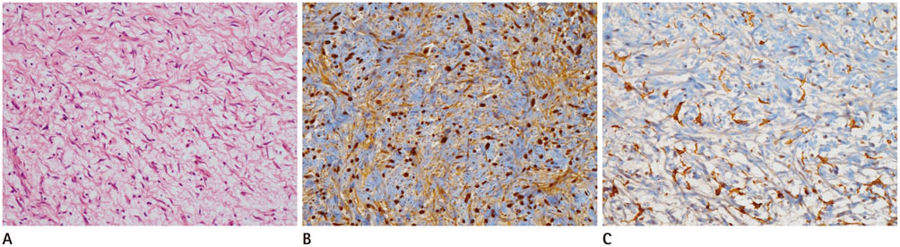
Fig. 3 Microscopic examination of the specimen. A. A microphotograph of the specimen shows evenly distributed spindle cells with wavy nuclei (hematoxylin-eosin stain, original magnification × 200). B, C. Immunochemical stain for S100 (B) and CD34 (C) show S100 positivity in most, but not all, cells (dark cells) and CD34 positivity in some stromal cells (original magnification × 200).
Reference
-
1. Fink D, Schneider C, Wight E, Perucchini D, Haller U. [Neurofibromatosis of the breast in a patient with Morbus von Recklinghausen]. Gynakol Geburtshilfliche Rundsch. 2000; 40:47–49.2. Sherman JE, Smith JW. Neurofibromas of the breast and nipple-areolar area. Ann Plast Surg. 1981; 7:302–307.3. Bongiorno MR, Doukaki S, Aricò M. Neurofibromatosis of the nipple-areolar area: a case series. J Med Case Rep. 2010; 4:22.4. Gokalp G, Hakyemez B, Kizilkaya E, Haholu A. Myxoid neurofibromas of the breast: mammographical, sonographical and MRI appearances. Br J Radiol. 2007; 80:e234–e237.5. Tucker T, Wolkenstein P, Revuz J, Zeller J, Friedman JM. Association between benign and malignant peripheral nerve sheath tumors in NF1. Neurology. 2005; 65:205–211.6. Sharif S, Moran A, Huson SM, Iddenden R, Shenton A, Howard E, et al. Women with neurofibromatosis 1 are at a moderately increased risk of developing breast cancer and should be considered for early screening. J Med Genet. 2007; 44:481–484.7. Millman SL, Mercado CL. An unusual presentation of neurofibromatosis of the breast. Breast J. 2004; 10:45–47.8. Lee SH, Park JM, Kook SH, Han BK, Moon WK. Metastatic tumors to the breast: mammographic and ultrasonographic findings. J Ultrasound Med. 2000; 19:257–262.9. Reynolds DL Jr, Jacobson JA, Inampudi P, Jamadar DA, Ebrahim FS, Hayes CW. Sonographic characteristics of peripheral nerve sheath tumors. AJR Am J Roentgenol. 2004; 182:741–744.10. Jeyaretna DS, Oriolowo A, Smith ME, Watkins RM. Solitary neurofibroma in the male breast. World J Surg Oncol. 2007; 5:23.
- Full Text Links
-
- Actions
-
Cited
- CITED
-
- Close
- Share
-
- Similar articles
-
- Random Synchronous Malignancy in Male Breast: A Case Report
- Occult Invasive Lobular Carcinoma of Breast Detected by Stomach Metastasis: A Case Report
- A Rare Case of Cardiac Neurofibroma in a Patient with Neurofibromatosis Type 1: Radiologic Findings
- A Case of Orbital Neurilemoma Associated with Neurofibroma tosis
- A Neurofibroma Confused with Sarcomatous Transformation on F-18 FDG PET/CT in Neurofibromatosis-1
