Endoscopic Fluorescence Angiography with Indocyanine Green : A Preclinical Study in the Swine
- Affiliations
-
- 1Department of Neurosurgery, Seoul National University Hospital, Seoul National University College of Medicine, Seoul, Korea.
- 2Department of Orthopaedic Surgery, Seoul National University Hospital, Seoul National University College of Medicine, Seoul, Korea.
- 3Department of Biomedical Engineering, Seoul National University Hospital, Seoul National University College of Medicine, Seoul, Korea.
- 4Korea Electrotechnology Research Institute Russia Science Seoul Center, Seoul, Korea. dslee@keri.re.kr
- KMID: 2151152
- DOI: http://doi.org/10.3340/jkns.2015.58.6.513
Abstract
OBJECTIVE
Microscopic indocyanine green (ICG) angiography is useful for identifying the completeness of aneurysm clipping and the preservation of parent arteries and small perforators. Neuroendoscopy is helpful for visualizing structures beyond the straight line of the microscopic view. We evaluated our prototype of endoscopic ICG fluorescence angiography in swine, which we developed in order to combine the merits of microscopic ICG angiography and endoscopy.
METHODS
Our endoscopic ICG system consists of a camera, a light source, a display and software. This system can simultaneously display real-time visible and near infrared fluorescence imaging on the same monitor. A commercially available endoscope was used, which was 4 mm in diameter and had an angle of 30degrees. A male crossbred swine was used.
RESULTS
Under general anesthesia, a small craniotomy was performed and the brain surface of the swine was exposed. ICG was injected via the ear vein with a bolus dose of 0.3 mg/kg. Visible and ICG fluorescence images of cortical vessels were simultaneously observed on the display monitor at high resolution. The real-time merging of the visible and fluorescent images corresponded well.
CONCLUSION
Simultaneous visible color and ICG fluorescent imaging of the cortical vessels in the swine brain was satisfactory. Technical improvement and clinical implication are expected.
MeSH Terms
Figure
-
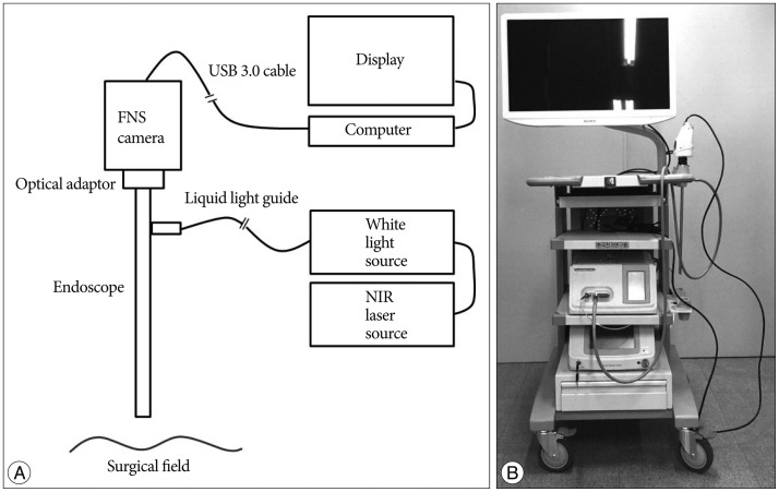
Fig. 1 Schematic illustration (A) and real model (B) of the endoscopic indocyanine green (ICG) fluorescence angiography.
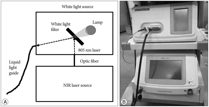
Fig. 2 Schematic illustration (A) and real model (B) of the white light and near infrared (NIR) laser sources.
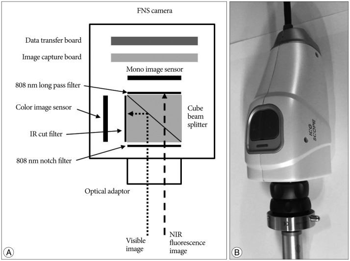
Fig. 3 Schematic illustration (A) and real model (B) of the fluorescence navigation surgery (FNS) camera.
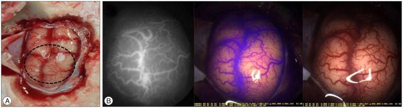
Fig. 4 Endoscopic visible color and ICG fluorescent images. A : Photography of the exposed brain cortex in the swine. Endoscopic imaging was targeted for the dotted circle area. B : ICG fluorescent image (left), visible color image (middle), and and real-time merging image of ICG fluorescent and visible color images (right).
Cited by 1 articles
-
Keyhole Approach and Neuroendoscopy for Cerebral Aneurysms
Won-Sang Cho, Jeong Eun Kim, Hyun-Seung Kang, Young-Je Son, Jae Seung Bang, Chang Wan Oh
J Korean Neurosurg Soc. 2017;60(3):275-281. doi: 10.3340/jkns.2017.0101.002.
Reference
-
1. Alexander TD, Macdonald RL, Weir B, Kowalczuk A. Intraoperative angiography in cerebral aneurysm surgery : a prospective study of 100 craniotomies. Neurosurgery. 1996; 39:10–17. discussion 17-18PMID: 8805135.2. Andaluz N, Zuccarello M. Recent trends in the treatment of cerebral aneurysms : analysis of a nationwide inpatient database. J Neurosurg. 2008; 108:1163–1169. PMID: 18518722.
Article3. Betz CS, Zhorzel S, Schachenmayr H, Stepp H, Havel M, Siedek V, et al. Endoscopic measurements of free-flap perfusion in the head and neck region using red-excited Indocyanine Green : preliminary results. J Plast Reconstr Aesthet Surg. 2009; 62:1602–1608. PMID: 19036663.
Article4. Brown RD Jr, Broderick JP. Unruptured intracranial aneurysms : epidemiology, natural history, management options, and familial screening. Lancet Neurol. 2014; 13:393–404. PMID: 24646873.
Article5. Bruneau M, Appelboom G, Rynkowski M, Van Cutsem N, Mine B, De Witte O. Endoscope-integrated ICG technology : first application during intracranial aneurysm surgery. Neurosurg Rev. 2013; 36:77–84. discussion 84-85PMID: 22918545.6. Ciamberlini C, Guarnieri V, Longobardi G, Poggi P, Donati MC, Panzardi G. Indocyanine green videoangiography using cooled charge-coupled devices in central serous choroidopathy. J Biomed Opt. 1997; 2:218–225. PMID: 23014876.
Article7. Fischer G, Oertel J, Perneczky A. Endoscopy in aneurysm surgery. Neurosurgery. 2012; 70(2 Suppl Operative):184–190. discussion 190-191PMID: 21937925.
Article8. Hernesniemi J, Ishii K, Niemelä M, Smrcka M, Kivipelto L, Fujiki M, et al. Lateral supraorbital approach as an alternative to the classical pterional approach. Acta Neurochir Suppl. 2005; 94:17–21. PMID: 16060236.
Article9. Ishihara R, Iishi H, Kidu T, Yamamoto S, Miyoshi R, Inoue T, et al. Infrared endoscopic system for bleeding-point detection after flushing with indocyanine green solution (with videos). Gastrointest Endosc. 2008; 68:975–981. PMID: 18984104.
Article10. Litvack ZN, Zada G, Laws ER Jr. Indocyanine green fluorescence endoscopy for visual differentiation of pituitary tumor from surrounding structures. J Neurosurg. 2012; 116:935–941. PMID: 22360574.
Article11. Mielke D, Malinova V, Rohde V. Comparison of intraoperative microscopic and endoscopic ICG angiography in aneurysm surgery. Neurosurgery. 2014; 10(Suppl 3):418–425. discussion 425PMID: 24618802.
Article12. Molyneux AJ, Kerr RS, Yu LM, Clarke M, Sneade M, Yarnold JA, et al. : International subarachnoid aneurysm trial (ISAT) of neurosurgical clipping versus endovascular coiling in 2143 patients with ruptured intracranial aneurysms : a randomised comparison of effects on survival, dependency, seizures, rebleeding, subgroups, and aneurysm occlusion. Lancet. 2005; 366:809–817. PMID: 16139655.
Article13. Nimura H, Narimiya N, Mitsumori N, Yamazaki Y, Yanaga K, Urashima M. Infrared ray electronic endoscopy combined with indocyanine green injection for detection of sentinel nodes of patients with gastric cancer. Br J Surg. 2004; 91:575–579. PMID: 15122608.
Article14. Nishiyama Y, Kinouchi H, Senbokuya N, Kato T, Kanemaru K, Yoshioka H, et al. Endoscopic indocyanine green video angiography in aneurysm surgery : an innovative method for intraoperative assessment of blood flow in vasculature hidden from microscopic view. J Neurosurg. 2012; 117:302–308. PMID: 22680246.
Article15. Park HS, Park SK, Han YM. Microsurgical experience with supraorbital keyhole operations on anterior circulation aneurysms. J Korean Neurosurg Soc. 2009; 46:103–108. PMID: 19763211.
Article16. Quiñones-Hinojosa A, Alam M, Lyon R, Yingling CD, Lawton MT. Transcranial motor evoked potentials during basilar artery aneurysm surgery : technique application for 30 consecutive patients. Neurosurgery. 2004; 54:916–924. discussion 924PMID: 15046658.
Article17. Raabe A, Beck J, Gerlach R, Zimmermann M, Seifert V. Near-infrared indocyanine green video angiography : a new method for intraoperative assessment of vascular flow. Neurosurgery. 2003; 52:132–139. discussion 139PMID: 12493110.
Article18. Raabe A, Nakaji P, Beck J, Kim LJ, Hsu FP, Kamerman JD, et al. Prospective evaluation of surgical microscope-integrated intraoperative near-infrared indocyanine green videoangiography during aneurysm surgery. J Neurosurg. 2005; 103:982–989. PMID: 16381184.
Article19. Reisch R, Perneczky A. Ten-year experience with the supraorbital subfrontal approach through an eyebrow skin incision. Neurosurgery. 2005; 57(4 Suppl):242–255. discussion 242-255PMID: 16234671.
Article20. Smith GA, Dagostino P, Maltenfort MG, Dumont AS, Ratliff JK. Geographic variation and regional trends in adoption of endovascular techniques for cerebral aneurysms. J Neurosurg. 2011; 114:1768–1777. PMID: 21314274.
Article
- Full Text Links
-
- Actions
-
Cited
- CITED
-
- Close
- Share
-
- Similar articles
-
- Utility of Indocyanine Green Fluorescence Imaging in Wound Assessment
- Indocyanine green angiography of retinal astrocytomas associated with tuberous sclerosis
- Fluorescein and Indocyanine Green Angiography of Choroidal Tumors
- Indocyanine Green-Guided Video-Assisted Thoracoscopic Surgery for Resection of an Ectopic Mediastinal Parathyroid Adenoma
- Availability of Optical Coherence Tomography in Diagnosis and Classification of Choroidal Neovascularization
