A Rare Case of Primary Tubular Adenocarcinoma of the Thymus, Enteric Immunophenotype: A Case Study and Review of the Literature
- Affiliations
-
- 1Department of Pathology, Soonchunhyang University Cheonan Hospital, Cheonan, Korea. mhoh0212@schmc.ac.kr
- 2Department of Pathology and Respiratory Center, Seoul National University Bundang Hospital, Seongnam, Korea.
- 3Division of Hematology and Oncology, Department of Internal Medicine, Soonchunhyang University Cheonan Hospital, Cheonan, Korea.
- KMID: 2151143
- DOI: http://doi.org/10.4132/jptm.2015.04.16
Abstract
- Thymic carcinomas are uncommon malignant tumors, and thymic adenocarcinomas are extremely rare. Here, we describe a case of primary thymic adenocarcinoma in a 59-year-old woman. Histological examination of the tumor revealed tubular morphology with expression of cytokeratin 20 and caudal-type homeobox 2 according to immunohistochemistry, suggesting enteric features. Extensive clinical and radiological studies excluded the possibility of an extrathymic primary tumor. A review of the literature revealed only two global cases of primary tubular adenocarcinomas of the thymus with enteric immunophenotype.
MeSH Terms
Figure
-
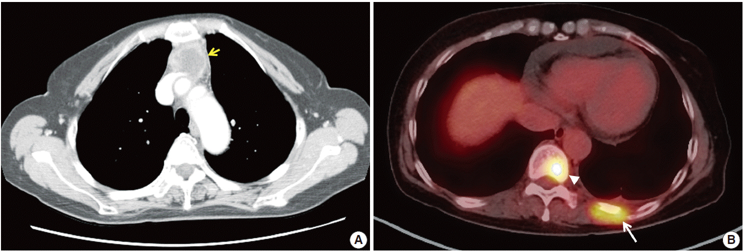
Fig. 1. Radiologic examination of the anterior mediastinum. (A) An irregularly enhancing mass (arrow) in the anterior mediastinum on a chest computed tomography scan. (B) Abnormal hyperuptake in lesions at the 10th vertebra (arrowhead) and the left 10th rib (arrow) on a wholebody positron emission tomography scan.
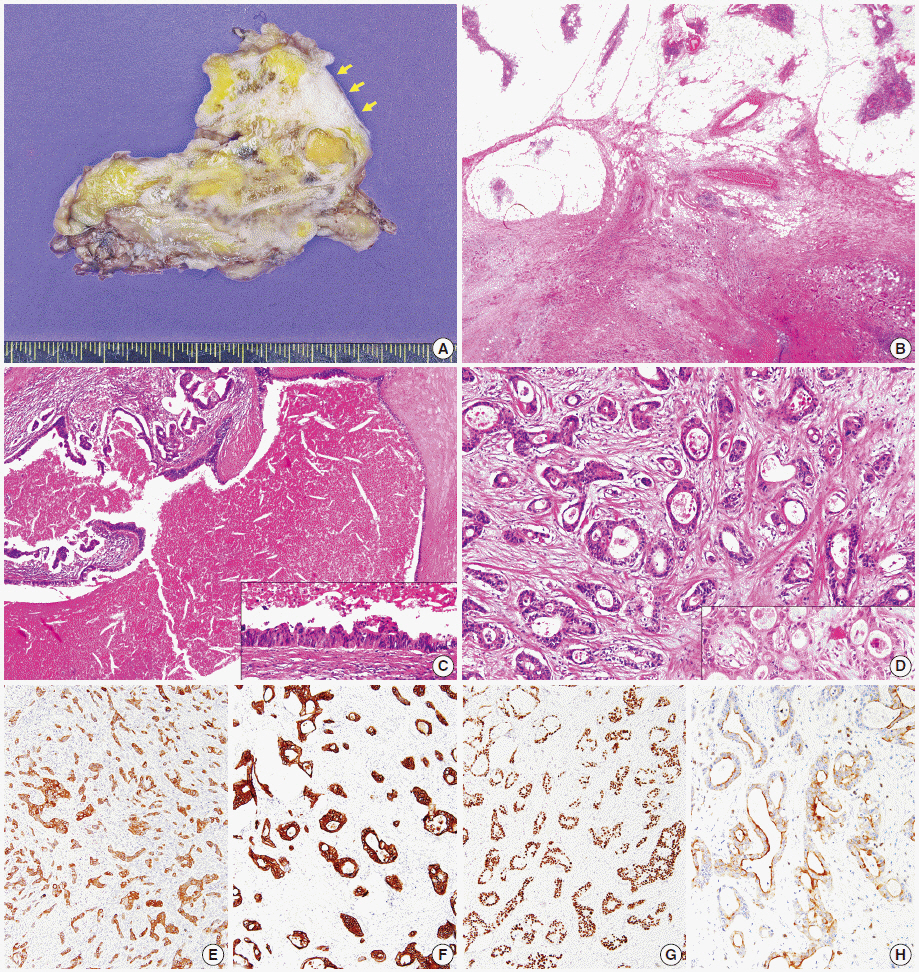
Fig. 2. Pathologic examination. (A) Gross examination of the tumor reveals an ill-defined mass with pericardial invasion (arrows, pericardium). (B) Microscopically, the tumor is surrounded by normal thymic tissues. At higher magnification, the tumor is composed of glandular or tubular structures with large glands lined by tall columnar cells (C) and oval cells forming small glands (D). The tumor cells show positive staining for CK7 (E), CK20 (F), CDX2 (G), and CD5 (H). CK, cytokeratin; CDX2, caudal type homeobox 2.
Cited by 1 articles
-
Cytologic Characteristics of Thymic Adenocarcinoma with Enteric Differentiation: A Study of Four Fine-Needle Aspiration Specimens
Ah-Young Kwon, Joungho Han, Hae-yon Cho, Seokhwi Kim, Heejin Bang, Jiyeon Hyeon
J Pathol Transl Med. 2017;51(5):509-512. doi: 10.4132/jptm.2017.03.22.
Reference
-
1. Müller-Hermelink HK, Marx A, Kuo TT, Kurrer M, Chen G, Shimosato Y. Non-papillary adenocarcinomas. In : Travis WD, Brambilla E, Müller-Hermelink HK, Harris CC, editors. World Health Organization classification of tumours: pathology and genetics of tumours of the lung, pleura, thymus and heart. 3rd ed. Lyon: IARC Press;2004. p. 184.2. Moriyama S, Shimizu N, Kurita A, Teramoto S, Taguchi K. A case of adenocarcinoma of the thymus. Nihon Kyobu Geka Gakkai Zasshi. 1989; 37:717–22.3. Maghbool M, Ramzi M, Nagel I, et al. Primary adenocarcinoma of the thymus: an immunohistochemical and molecular study with review of the literature. BMC Clin Pathol. 2013; 13:17.
Article4. Matsuno Y, Morozumi N, Hirohashi S, Shimosato Y, Rosai J. Papillary carcinoma of the thymus: report of four cases of a new microscopic type of thymic carcinoma. Am J Surg Pathol. 1998; 22:873–80.5. Yoshino M, Hiroshima K, Motohashi S, et al. Papillary carcinoma of the thymus gland. Ann Thorac Surg. 2005; 80:741–2.
Article6. Furtado A, Nogueira R, Ferreira D, Tente D, Eisele R, Parente B. Papillary adenocarcinoma of the thymus: case report and review of the literature. Int J Surg Pathol. 2010; 18:530–3.
Article7. Choi WW, Lui YH, Lau WH, Crowley P, Khan A, Chan JK. Adenocarcinoma of the thymus: report of two cases, including a previously undescribed mucinous subtype. Am J Surg Pathol. 2003; 27:124–30.8. Ra SH, Fishbein MC, Baruch-Oren T, et al. Mucinous adenocarcinomas of the thymus: report of 2 cases and review of the literature. Am J Surg Pathol. 2007; 31:1330–6.9. Abdul-Ghafar J, Yong SJ, Kwon W, Park IH, Jung SH. Primary thymic mucinous adenocarcinoma: a case report. Korean J Pathol. 2012; 46:377–81.
Article10. Sawai T, Inoue Y, Doi S, et al. Tubular adenocarcinoma of the thymus: case report and review of the literature. Int J Surg Pathol. 2006; 14:243–6.
Article11. Misao T, Yamamoto Y, Nakano H, Toyooka S, Yamane M, Satoh K. Primary thymic adenocarcinoma with production of carbohydrate antigen 19-9 and carcinoembryonic antigen. Jpn J Thorac Cardiovasc Surg. 2004; 52:30–2.
Article12. Moser B, Schiefer AI, Janik S, et al. Adenocarcinoma of the thymus, enteric type: report of 2 cases, and proposal for a novel subtype of thymic carcinoma. Am J Surg Pathol. 2015; 39:541–8.13. Ishiwata T, Sekiya M, Suzuki T, Matsuoka T, Kumasaka T, Takahashi K. Thymic adenocarcinoma with sarcomatoid features characterized by intracaval tumor growth: report of a case. Surg Today. 2010; 40:1068–72.
Article14. Teramoto K, Kawaguchi Y, Hori T, et al. Thymic papillo-tubular adenocarcinoma containing a cyst: report of a case. Surg Today. 2012; 42:988–91.
Article15. Suster S, Rosai J. Thymic carcinoma: a clinicopathologic study of 60 cases. Cancer. 1991; 67:1025–32.
Article16. Wang S, Wang Z, Liu X, Wang D, Liu F. Prognostic factors of patients with thymic carcinoma after surgery: a retrospective analysis of 58 cases. World J Surg. 2014; 38:2032–8.
Article17. Filosso PL, Guerrera F, Rendina AE, et al. Outcome of surgically resected thymic carcinoma: a multicenter experience. Lung Cancer. 2014; 83:205–10.
Article
- Full Text Links
-
- Actions
-
Cited
- CITED
-
- Close
- Share
-
- Similar articles
-
- Cytologic Characteristics of Thymic Adenocarcinoma with Enteric Differentiation: A Study of Four Fine-Needle Aspiration Specimens
- Mucinous Tubular and Spindle Cell Carcinoma of Kidney Occurring in a Patient with Pulmonary Adenocarcinoma
- A Case of Tubular Adenocarcinoma on Fistula of Duodenal Bulb
- A case of true thymic hyperplasia in the mediastinum with ectopic thymus in the neck
- A Case of Malignant Transformation of Gastric Tubular Adenoma Proven by 9-year Follow-Up
