Absence of the Septum Pellucidum Associated with a Midline Fornical Nodule and Ventriculomegaly: A Report of Two Cases
- Affiliations
-
- 1Department of Pathology, Cheil General Hospital and Women's Healthcare Center, Kwandong University College of Medicine, Seoul, Korea. ykcmd@naver.com
- 2Department of Pathology, Seoul National University College of Medicine, Seoul, Korea.
- KMID: 2150883
- DOI: http://doi.org/10.3346/jkms.2010.25.6.970
Abstract
- We report two autopsy cases that revealed the partial absence of the septum pellucidum with ventriculomegaly. In each case, the brain showed mild dilatation of both frontal horns of the lateral ventricles, normal third and fourth ventricles and no aqueductal stenosis. The posterior portion of the septum pellucidum was absent and the fornices were fused in a single midline nodule, abnormally displaced to a caudal position and lodged in the foramina of Monro. The brain base showed no apparent abnormalities; the optic nerves were well developed. We conclude that the caudally displaced fornix in the absence of the septum pellucidum may have intermittently obstructed the foramina of Monro and induced mild ventriculomegaly.
Figure
-
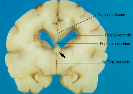
Fig. 1 Coronal section of the Case 1 brain through the interventricular foramen. The lateral ventricles are mildly distended and the third ventricle is normal. A single midline fornical nodule (arrow) almost totally obstructs the foramina of Monro. The septum pellucidum is absent in its posterior portion and the corpus callosum seems normal.
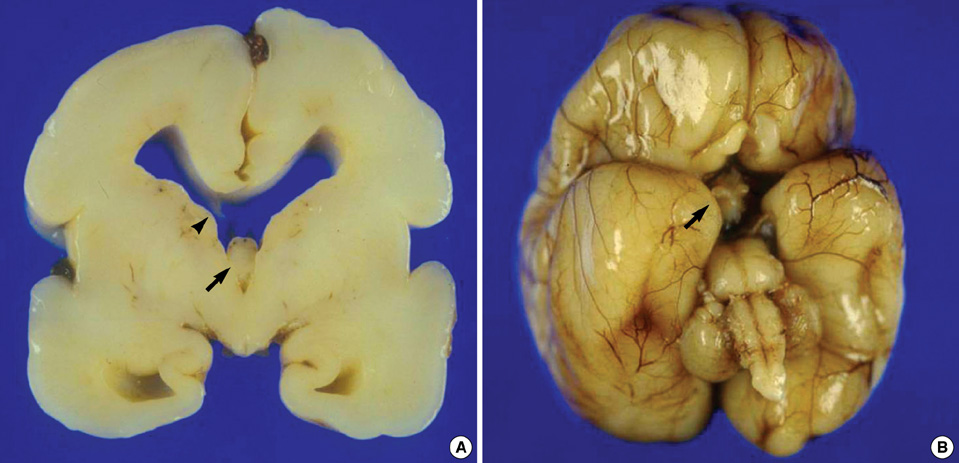
Fig. 2 Gross findings of the brain of the Case 2. (A) Coronal section through the interventricular foramen. The frontal horns of the lateral ventricles are mildly distended with blunt angles. A single midline fornical nodule (arrow) obstructs the foramina of Monro. Wisps of the anterior part of the septum pellucidum are seen (arrow head). An iatrogenic tear of the corpus callosum is present. (B) The base of the brain shows well developed optic nerves (arrow), olfactory nerves and other cranial nerves. Gyri and sulci show no gross abnormalities.
Reference
-
1. Bruyn GW. Vinken PJ, Bruyn GW, editors. Agenesis septi pellucidi, cavum septi pellucidi, cavum vergae, and cavum veli interpositi. Handbook of clinical neurology, vol. 30. Congenital malformations of the brain and skull. Part I. 1977. Amsterdam: North Holland;299–336.2. Barkovich AJ, Norman D. Absence of the septum pellucidum: a useful sign in the diagnosis of congenital brain malformations. AJR Am J Roentgenol. 1989. 152:353–360.3. Scoffings DJ, Kurian KM. Congenital and acquired lesions of the septum pellucidum. Clin Radiol. 2008. 63:210–219.4. Lepinard C, Coutant R, Boussion F, Loisel D, Delorme B, Biquard F, Bonneau D, Guichet A, Descamps P. Prenatal diagnosis of absence of the septum pellucidum associated with septo-optic dysplasia. Ultrasound Obstet Gynecol. 2005. 25:73–75.5. Willnow S, Kiess W, Butenandt O, Dorr HG, Enders A, Strasser-Vogel B, Egger J, Schwarz HP. Endocrine disorders in septo-optic dysplasia (De Morsier syndrome)-evaluation and follow up of 18 patients. Eur J Pediatr. 1996. 155:179–184.6. Polizzi A, Pavone P, Iannetti P, Manfré L, Ruggieri M. Septo-optic dysplasia complex: a heterogeneous malformation syndrome. Pediatr Neurol. 2006. 34:66–71.7. Belhocine O, André C, Kalifa G, Adamsbaum C. Does asymptomatic septal agenesis exist? A review of 34 cases. Pediatr Radiol. 2005. 35:410–418.8. Malinger G, Lev D, Kidron D, Heredia F, Hershkovitz R, Lerman-Sagie T. Differential diagnosis in fetuses with absent septum pellucidum. Ultrasound Obstet Gynecol. 2005. 25:42–49.9. Celentano C, Prefumo F, Liberati M, Tartaro A, Gallo G, Lattanzio G, Rotmensch S. Prenatal diagnosis of septal agenesis with normal pituitary function. Prenat Diagn. 2006. 26:1075–1077.10. Marshall I, Ugrasbul F, Manginello F, Wajnrajch MP, Shackleton CH, New MI, Vogiatzi MV. Congenital hypopituitarism as a cause of undetectable estriol levels in the maternal triple-marker screen. J Clin Endocrinol Metab. 2003. 88:4144–4148.11. Koo H, Lee KY, Chi JG. Congenital hydrocephalus associated with anomalies of midline telencephalic structures. Pathol Res Pract. 1991. 187:939–942.
