A New Reporter Vector System Based on Flow-Cytometry to Detect Promoter Activity
- Affiliations
-
- 1School of Life Sciences and Biotechnology, Korea University, Seoul 136-701, Korea. sehopark@korea.ac.kr
- 2Department of Microbiology, Gachon Medical School, Inchon 405-760, Korea.
- KMID: 2150652
- DOI: http://doi.org/10.4110/in.2009.9.6.243
Abstract
- In this study, we report the development of a new dual reporter vector system for the analysis of promoter activity. This system employs green fluorescence emitting protein, EGFP, as a reporter, and uses red fluorescence emitting protein, DsRed, as a transfection control in a single vector. The expression of those two proteins can be readily detected via flow cytometry in a single analysis, with no need for any further manipulation after transfection. As this system allows for the simultaneous detection of both the control and reporter proteins in the same cells, only transfected cells which express the control protein, DsRed, can be subjected to promoter activity analysis, via the gating out of all un-transfected cells. This results in a dramatic increase in the promoter activity detection sensitivity. This novel reporter vector system should prove to be a simple and efficient method for the analysis of promoter activity.
MeSH Terms
Figure
-
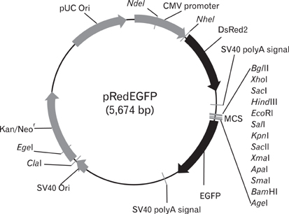
Figure 1 Construction of the dual reporter vector, pRedEGFP. The CMV promoter-driven DsRed gene, which is expressed constitutively in most mammalian cells, and the promoter-less EGFP gene are delineated within a single vector. The region upstream of EGFP includes multicloning sites for promoter cloning.

Figure 2 Expression of EGFP in DsRed-positive cells. (A) Promoter regions of mouse CD1d1 were cloned into the MCS of pRedEGFP and transfected into the Bcl-1 lymphoma cell line. Cells were harvested 72 hours after transfection. (B, C) In flow cytometric analyses, transfected cells, which evidence constitutive DsRed expression (above the horizontal line of each panel), were gated (B), and their EGFP expression is presented in histograms (C). EGFP expression was readily detectable among the DsRed-positive population, although the transfection efficiency was rather low (~5%, the ratio of DsRed-positive cells). The TA (transcription activity) of the DsRed-positive cells was calculated as follows: (MFI of EGFP/MFI of DsRed)×100%. The X and Y-axes of the dot-plot and the X-axis of the histogram-plot were represented on a log scale, from 100 to 104. The Y-axis of histogram-plot is shown as a linear scale.

Figure 3 Comparison of the dual reporter system with the conventional CAT reporter system. The same promoter regions used in the dual reporter vector system were re-cloned into the CAT reporter system, pCAT®3-Enhancer vector (A). Vectors harboring different lengths of the promoter region were cotransfected with pSV-β-Galactosidase vector. The transcriptional activities of each region were determined via Chloramphenicol acetyl transferase activity assays (B, C) after compensation with beta-galactosidase. The pCAT3 control vector without the promoter sequence (pCAT®3-Basic) and the one harboring the SV40 promoter (pCAT®3-Control) were used as negative and positive controls, respectively.
Reference
-
1. Dominguez P, Ibaraki K, Robey PG, Hefferan TE, Termine JD, Young MF. Expression of the osteonectin gene potentially controlled by multiple cis- and trans-acting factors in cultured bone cells. J Bone Miner Res. 1991. 6:1127–1136.
Article2. Gorman CM, Moffat LF, Howard BH. Recombinant genomes which express chloramphenicol acetyltransferase in mammalian cells. Mol Cell Biol. 1982. 2:1044–1051.
Article3. Alam J, Cook JL. Reporter genes: application to the study of mammalian gene transcription. Anal Biochem. 1990. 188:245–254.
Article4. Russo-Marie F, Roederer M, Sager B, Herzenberg LA, Kaiser D. Beta-galactosidase activity in single differentiating bacterial cells. Proc Natl Acad Sci U S A. 1993. 90:8194–8198.
Article5. Dietrich C, Maiss E. Red fluorescent protein DsRed from Discosoma sp. as a reporter protein in higher plants. Biotechniques. 2002. 32:286–293.
Article6. Nancharaiah YV, Wattiau P, Wuertz S, Bathe S, Mohan SV, Wilderer PA, Hausner M. Dual labeling of Pseudomonas putida with fluorescent proteins for in situ monitoring of conjugal transfer of the TOL plasmid. Appl Environ Microbiol. 2003. 69:4846–4852.
Article7. Cormack BP, Valdivia RH, Falkow S. FACS-optimized mutants of the green fluorescent protein (GFP). Gene. 1996. 173(1 Spec No):33–38.
Article8. Gross LA, Baird GS, Hoffman RC, Baldridge KK, Tsien RY. The structure of the chromophore within DsRed, a red fluorescent protein from coral. Proc Natl Acad Sci U S A. 2000. 97:11990–11995.
Article9. Davey HM, Kell DB. Flow cytometry and cell sorting of heterogeneous microbial populations: the importance of single-cell analyses. Microbiol Rev. 1996. 60:641–696.
Article
- Full Text Links
-
- Actions
-
Cited
- CITED
-
- Close
- Share
-
- Similar articles
-
- Heterologous Regulation of BCG hsp65 Promoter by M. leprae 18 kDa Transcription Repression Responsive Element
- Development of an efficient endothelial cell specific vector using promoter and 5' untranslated sequences from the human preproendothelin-1 gene
- Recombinant AAV Vector with MITF-M Promoter for Melanoma Gene Therapy
- Analysis of T Cells Using Flow Cytometry
- A Novel Rapid Fungal Promoter Analysis System Using the Phosphopantetheinyl Transferase Gene, npgA, in Aspergillus nidulans
