Multifocal Sparganosis Mimicking Lymphoma Involvement: Multimodal Imaging Findings of Ultrasonography, CT, MRI, and Positron Emission Tomography-Computed Tomography
- Affiliations
-
- 1Department of Radiology, Myongji Hospital, Seonam University College of Medicine, Goyang, Korea. zzzz3@hanmail.net
- 2Department of Pathology, Myongji Hospital, Seonam University College of Medicine, Goyang, Korea.
- 3Division of Hematology-Oncology, Department of Internal Medicine, Inje University Ilsan Paik Hospital, Goyang, Korea.
- 4Division of Hematology-Oncology, Department of Internal Medicine, Seoul Medical Center, Seoul, Korea.
- KMID: 2150463
- DOI: http://doi.org/10.3348/jksr.2016.74.1.55
Abstract
- Sparganosis is a rare parasitic disease caused by the migrating plerocercoid larva of Spirometra species tapeworms. The most frequent clinical manifestation is a subcutaneous nodule resembling a neoplasm. In this study, we presented multimodal findings of ultrasonography, computed tomography, magnetic resonance imaging, positron emission tomography-computed tomography and follow-up imagings on multifocal sparganosis, mimicking lymphoma involvement in a patient with lymphoma.
MeSH Terms
Figure
-
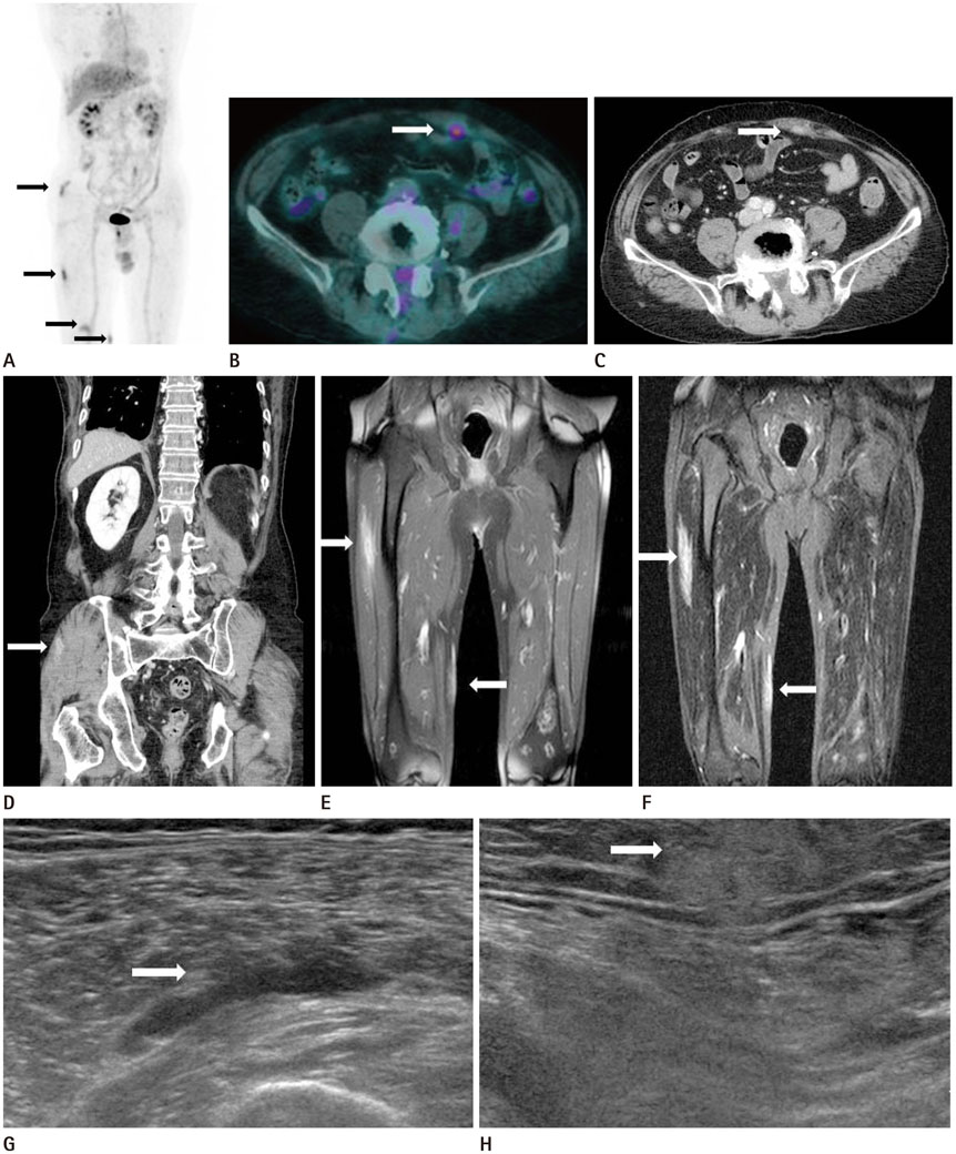
Fig. 1 PET-CT, abdomen CT, femur MRI and US images after 3 cycles of R-CHOP. A, B. PET-CT reveals multiple hypermetabolic lesions (SUV = 3.6-5.2) with tubular shape in the right buttock, vastus lateralis muscle, medial thigh (A, black arrows) and left rectus abdominis (B, white arrow). C, D. Abdomen CT shows ill-defined peripheral rim enhancing lesions with central low attenuation in the left rectus abdominis (C, white arrow) and right gluteus muscle (D, white arrow). E, F. Femur MRI shows peripherally enhancing tubular lesion in the right vastus lateralis muscle and homogeneous enhancing elongated lesion in the subcutaneous layer of the right medial thigh (E, white arrows). The lesions shows high signal intensity on STIR image (F, white arrows). G, H. US reveals a hypoechoic lesion with tubular shape in the right vastus lateralis (G, white arrow) and a heterogeneous hyperechoic lesion with ill-defined margin in the subcutaneous layer of the medial thigh (H, white arrow). CT = computed tomography, MRI = magnetic resonance imaging, PET-CT = positron emission tomography-computed tomography, R-CHOP = rituximab, cyclophosphamide, hydroxydaunorubicin (doxorubicin), oncovin (vincristine), prednisone, STIR = short tau inversion recovery, SUV = standardized uptake value, US = ultrasonography
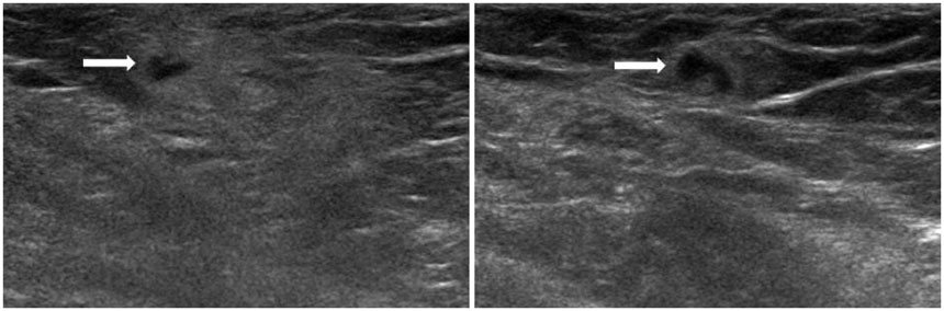
Fig. 2 Follow-up US after 2 weeks. US reveals a new, approximately 2.7 × 1.2 cm ill-defined hyperechoic lesion with internal small tubular hypoechoic area (white arrows) in the subcutaneous layer of left perineum. US = ultrasonography

Fig. 3 Histopathologic examination of the right medial thigh. A. The sparganum shows eosinophilic folded tegument, subtegumental muscle fibers and calcospherules (hematoxylin-eosin, × 40). B. Higher magnification of the sparganum in A. The tegument is thick and muscle fibers are oriented longitudinally (hematoxylin-eosin, × 100).
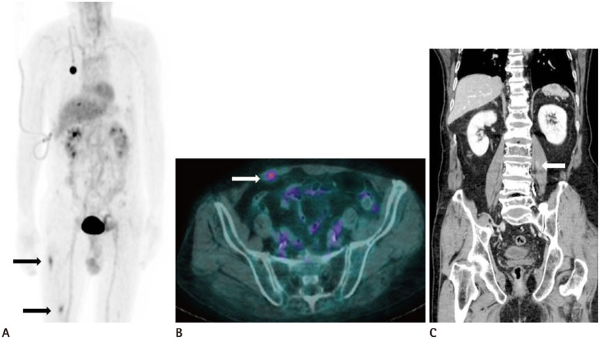
Fig. 4 Follow-up PET-CT after 2 months and abdomen CT after 9 months. A, B. On PET-CT, a hypermetabolic tubular lesion in right buttock has disappeared, but the other lesions in right lateral thigh (A, black arrows) still remains (A) and a hypermetabolic lesion has newly developed in the right rectus abdominis muscle (B, white arrow). C. Abdomen CT shows a new peripherally enhancing tubular lesion in the left psoas muscle (white arrow). CT = computed tomography, PET-CT = positron emission tomography-computed tomography
Reference
-
1. Lee BJ, Ahn SK, Kim SC, Lee SH. Clinical and histopathologic study of sparganosis. Korean J Dermatol. 1992; 30:168–174.2. Roh SY, Lee JY, Park KW, Jung SN. Sparganosis in a patient with diffuse large B cell lymphoma. J Cancer Res Ther. 2013; 9:712–714.3. Lee EK, Yoo YB. Axillary sparganosis which was misunderstood lymph node metastasis during neoadjuvant chemotheraphy in a breast cancer patient. Ann Surg Treat Res. 2014; 87:336–339.4. Kim JI, Kim TW, Hong SM, Moon TY, Lee IS, Choi KU, et al. Intramuscular sparganosis in the gastrocnemius muscle: a case report. Korean J Parasitol. 2014; 52:69–73.5. Park HJ, Park NH, Lee EJ, Park CS, Lee SM, Park SI. Ultrasonographic Findings of Subcutaneous and Muscular Sparganosis. J Korean Soc Radiol. 2009; 61:183–187.6. Hung GD, Chen YH, Chen DY, Lan JL. Subcutaneous panniculitis-like T-cell lymphoma presenting with hemophagocytic lymphohistiocytosis and skin lesions with characteristic high-resolution ultrasonographic findings. Clin Rheumatol. 2007; 26:775–778.7. Kim YH, Kim HS, Kim SY, Hwang YJ, Seo JW, Lee JY, et al. MR findings of subcutaneous panniculitis-like T-cell lymphoma: a case report. J Korean Radiol Soc. 2007; 57:479–482.8. Song T, Wang WS, Zhou BR, Mai WW, Li ZZ, Guo HC, et al. CT and MR characteristics of cerebral sparganosis. AJNR Am J Neuroradiol. 2007; 28:1700–1705.9. Tsai MD, Chang CN, Ho YS, Wang AD. Cerebral sparganosis diagnosed and treated with stereotactic techniques. Report of two cases. J Neurosurg. 1993; 78:129–113.
- Full Text Links
-
- Actions
-
Cited
- CITED
-
- Close
- Share
-
- Similar articles
-
- Diffuse Large B-Cell Lymphoma in the Era of Precision Oncology: How Imaging Is Helpful
- Follicular Lymphoma mimicking Metastatic Nodes on the F-18 FDG PET/CT and MRI for Staging of Endometrial Cancer
- CT, Magnetic Resonance, and 18F-Fluorodeoxyglucose Positron Emission Tomography/CT Imaging Features of Mucosa-Associated Lymphoid Tissue Lymphoma Involving Medial Rectus Muscle: A Case Report
- Positron Emission Tomography-CT, CT, and MR Imaging Findings of Tumor-Mimicking Organized Hematoma in the Maxillary Sinus: Two Case Reports
- A Case of Recurrence-Mimicking Charcoal Granuloma in a Breast Cancer Patient: Ultrasound, CT, PET/CT and Breast-Specific Gamma Imaging Findings
