A Case of Recurrence-Mimicking Charcoal Granuloma in a Breast Cancer Patient: Ultrasound, CT, PET/CT and Breast-Specific Gamma Imaging Findings
- Affiliations
-
- 1Department of Radiology, Myongji Hospital, Seonam University College of Medicine, Goyang, Korea. zzzz3@hanmail.net
- 2Department of General Surgery, Myongji Hospital, Seonam University College of Medicine, Goyang, Korea.
- 3Department of Pathology, Myongji Hospital, Seonam University College of Medicine, Goyang, Korea.
- 4Division of Hematology-Oncology, Department of Internal Medicine, Inje University Ilsan Paik Hospital, Goyang, Korea.
- KMID: 2327368
- DOI: http://doi.org/10.3348/jksr.2016.75.1.57
Abstract
- Charcoal remains stable without causing a foreign body reaction and it may be used for preoperative localization of a non-palpable breast mass. However, cases of post-charcoal-marking granuloma have only rarely been reported in the breast, and a charcoal granuloma can be misdiagnosed as malignancy. Herein, we report the ultrasound, computed tomography (CT), 18F-fluorodeoxyglucose-positron emission tomography/CT, and breast-specific gamma imaging findings of recurrence-mimicking charcoal granuloma after breast conserving surgery, following localization with charcoal in a breast cancer patient.
MeSH Terms
Figure
-
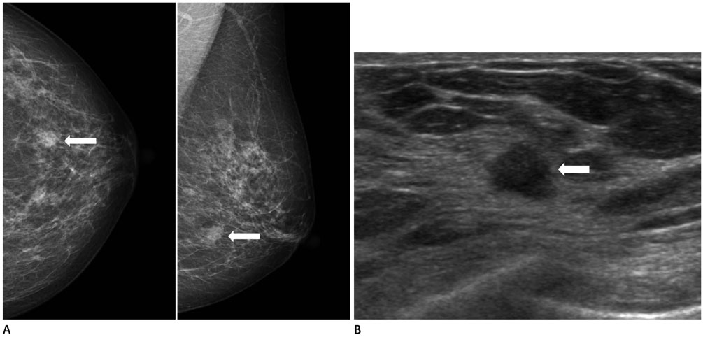
Fig. 1 Preoperative mammography and breast ultrasound of a 60-year-old woman. A. Mammography reveals a 0.9 cm mass (arrows) with an indistinct margin in the left lower outer quadrant of the breast. B. Breast ultrasound shows a 0.8 cm hypoechoic mass (arrow) with an indistinct margin and a subtle irregular shape at the 4 o'clock position in the left breast.
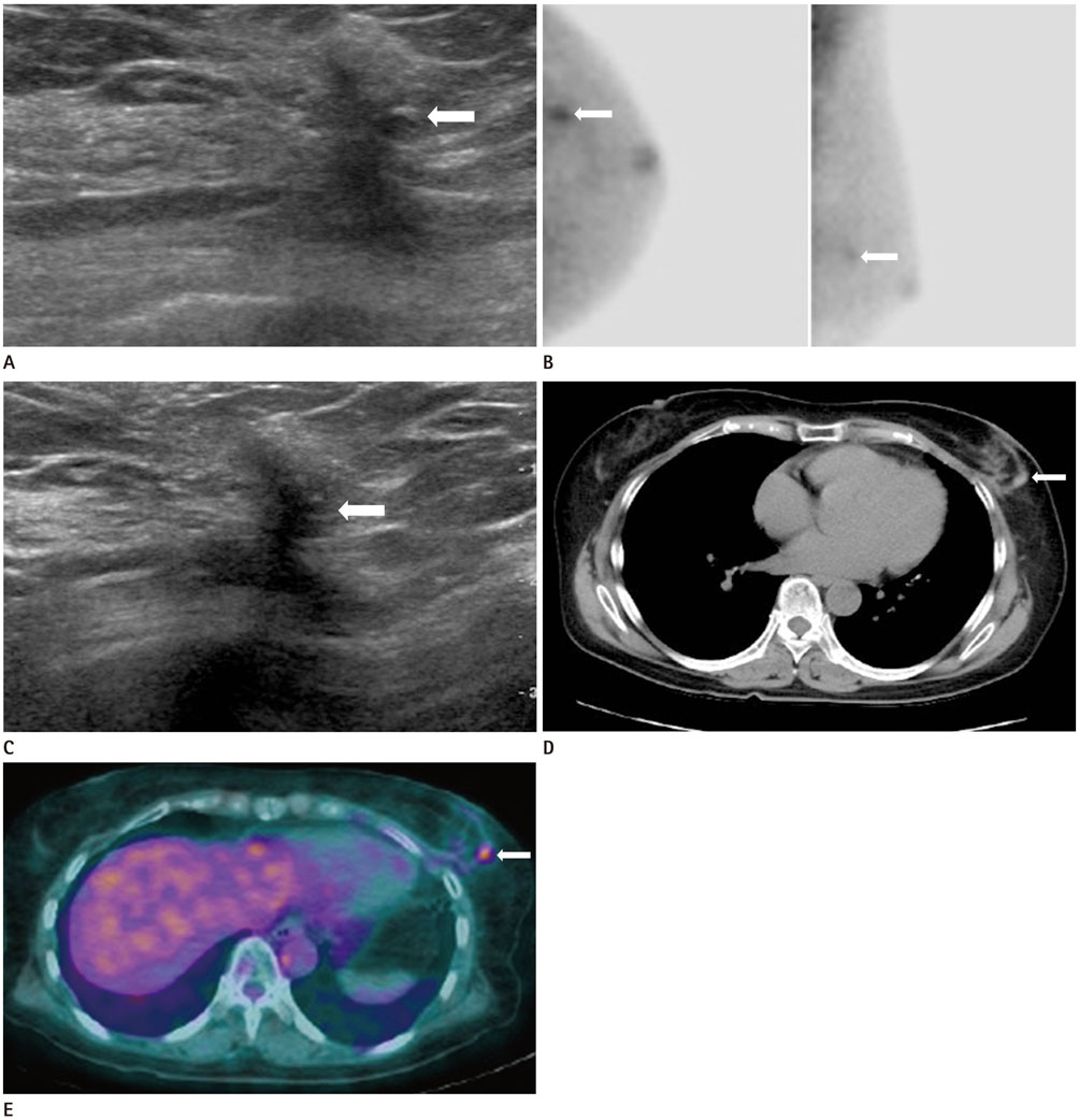
Fig. 2 BSGI, US, FDG-PET/CT, and chest CT findings of a charcoal granuloma. A. The first 6-month follow-up US after surgery demonstrates a spiculated irregular hypoechoic lesion (arrow) in the left middle outer quadrant of the breast. B. The first BSGI reveals intense focal uptake (arrows) in the left middle outer quadrant of the breast and it was considered as a postoperative change or recurrent lesion. C. The second 12-month follow-up US after surgery shows no significant change in the spiculated irregular hypoechoic lesion (arrow) in the left breast. D. Chest CT shows that the lesion is an ovoid hyperdense mass (47–66 Hounsfield units) (arrow) with adjacent postoperative distortion and it is not significantly enhanced after contrast administration. E. FDG-PET/CT shows a hypermetabolic lesion (arrow) (SUVmax = 2.6–3.7) in the previous operative bed of the left middle outer quadrant of the breast. BSGI = breast-specific gamma imaging, FDG-PET/CT = 18F-fluorodeoxyglucose-positron emission tomography/computed tomography, SUVmax = maximum standardized uptake value, US = ultrasound

Fig. 3 Histopathological examination of the charcoal granuloma. A. Gross specimen is dark-pigmented soft tissue fragments. B. Several multinucleated giant cells with adjoining pigmented material (arrow) (H&E, × 600) are evident. H&E = hematoxylin and eosin
Reference
-
1. Langlois SL, Carter ML. Carbon localisation of impalpable mammographic abnormalities. Australas Radiol. 1991; 35:237–241.2. Patrikeos A, Wylie EJ, Bourke A, Frost F. Imaging of carbon granulomas of the breast following carbon track localization. Clin Radiol. 1998; 53:845–848.3. Ruiz-Delgado ML, López-Ruiz JA, Sáiz-López A. Abnormal mammography and sonography associated with foreign-body giant-cell reaction after stereotactic vacuum-assisted breast biopsy with carbon marking. Acta Radiol. 2008; 49:1112–1118.4. Kim YK, Park HS. Foreign body granuloma of activated charcoal. Abdom Imaging. 2008; 33:94–97.5. Lim ST, Jeong HJ, Kim DW, Yim CY, Sohn MH. F-18 FDG PET-CT findings of intraperitoneal carbon particles-induced granulomas mimicking peritoneal carcinomatosis. Clin Nucl Med. 2008; 33:321–324.6. Choi JW, Moon WJ, Choi N, Roh HG, Kim MY, Kim NR, et al. Charcoal-induced granuloma that mimicked a nodal metastasis on ultrasonography and FDG-PET/CT after neck dissection. Korean J Radiol. 2015; 16:196–200.7. Brem RF, Floerke AC, Rapelyea JA, Teal C, Kelly T, Mathur V. Breast-specific gamma imaging as an adjunct imaging modality for the diagnosis of breast cancer. Radiology. 2008; 247:651–657.8. Zhou M, Johnson N, Gruner S, Ecklund GW, Meunier P, Bryn S, et al. Clinical utility of breast-specific gamma imaging for evaluating disease extent in the newly diagnosed breast cancer patient. Am J Surg. 2009; 197:159–163.9. Goldsmith SJ, Parsons W, Guiberteau MJ, Stern LH, Lanzkowsky L, Weigert J. SNM practice guideline for breast scintigraphy with breast-specific gamma-cameras 1.0. J Nucl Med Technol. 2010; 38:219–224.10. Bonhomme-Faivre L, Depraetere P, Savelli MP, Amdidouche D, Bizi E, Seiller M, et al. Charcoal suspension for tumor labelling modifies macrophage activity in mice. Life Sci. 2000; 66:817–827.
- Full Text Links
-
- Actions
-
Cited
- CITED
-
- Close
- Share
-
- Similar articles
-
- False Positive of F-18 FDG-PET/CT due to Activated Charcoal Granuloma from Intraperitoneal Chemotherapy: A Case Report
- [11C]Choline PET/CT in a Patient with Prostate Cancer Biochemical Recurrence Showing Two Suspicious Findings in the Breast and Liver
- Unusual Horner's Syndrome in Recurrent Breast Cancer: Evaluation Using ¹â¸F-FDG PET/CT
- Charcoal-Induced Granuloma That Mimicked a Nodal Metastasis on Ultrasonography and FDG-PET/CT after Neck Dissection
- COVID-19 Vaccine-Related Axillary and Cervical Lymphadenopathy in Patients with Current or Prior Breast Cancer and Other Malignancies: Cross-Sectional Imaging Findings on MRI, CT, and PET-CT
