Use of cone-beam computed tomography and three-dimensional modeling for assessment of anomalous pulp canal configuration: a case report
- Affiliations
-
- 1Department of Oral and Maxillofacial Radiology, Faculty of Dentistry, Kocaeli University, Kocaeli, Turkey. alpersinanoglu@yahoo.com
- 2Department of Endodontics, Faculty of Dentistry, Kocaeli University, Kocaeli, Turkey.
- 3Department of Biomechanical Engineering, Faculty of Technology, Kocaeli University, Kocaeli, Turkey.
- KMID: 2148867
- DOI: http://doi.org/10.5395/rde.2015.40.2.161
Abstract
- Three-dimensional (3D) reconstruction of cone-beam computed tomography (CBCT) scans appears to be a valuable method for assessing pulp canal configuration. The aim of this report is to describe endodontic treatment of a mandibular second premolar with aberrant pulp canal morphology detected by CBCT and confirmed by 3D modeling. An accessory canal was suspected during endodontic treatment of the mandibular left second premolar in a 21 year-old woman with a chief complaint of pulsating pain. Axial cross-sectional CBCT scans revealed that the pulp canal divided into mesiobuccal, lingual, and buccal canals in the middle third and ended as four separate foramina. 3D modeling confirmed the anomalous configuration of the fused root with a deep lingual groove. Endodontic treatment of the tooth was completed in two appointments. The root canals were obturated using lateral compaction of gutta-percha and root canal sealer. The tooth remained asymptomatic and did not develop periapical pathology until 12 months postoperatively. CBCT and 3D modeling enable preoperative evaluation of aberrant root canal systems and facilitate endodontic treatment.
Keyword
MeSH Terms
Figure
-

Figure 1 (a) Pre-operative periapical radiograph; (b) Intra-operative periapical radiograph; (c) Post-operative periapical radiograph; (d) 12 months follow-up periapical radiograph of the case.
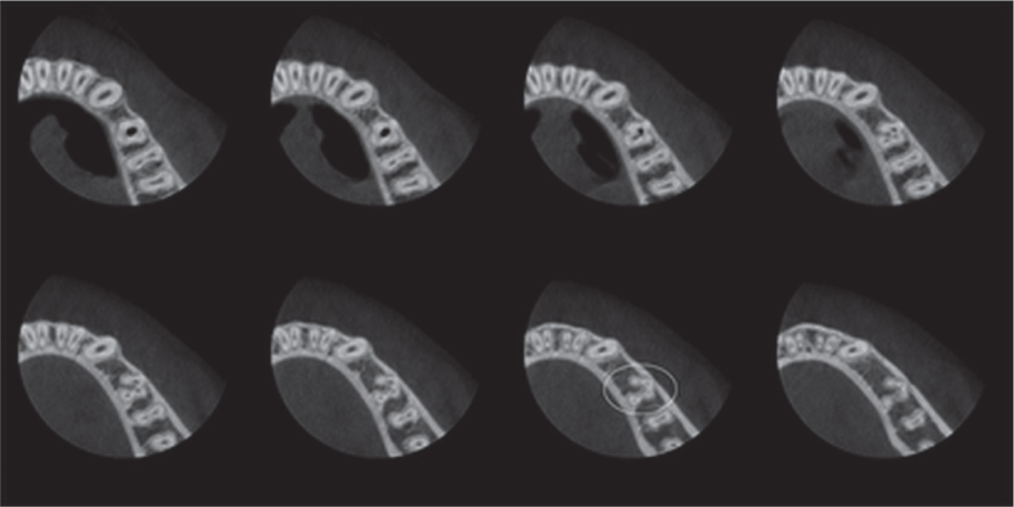
Figure 2 Axial sectional images revealed by CBCT. Note the 4 separate canal configurations marked by circle.

Figure 3 (a) Sagittal image of the root; (b) Axial image at the apical third presenting a C-shaped canal configuration; (c) Cross-sectional three-dimensional reconstruction demonstrating 4 separate canals.
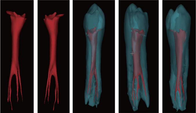
Figure 4 Three-dimensional modeling of the internal and external root canal morphology. Internal view (red) shows the separate root canal configuration, and the external view (blue) presents fused formation of the anomalous root.
Reference
-
1. Directorate-General for Energy Directorate D-Nuclear Energy Unit D4-Radiation Protection: Radiation protection no. 172: cone beam CT for dental and maxillofacial radiology, Evidence Based Guidelines. Luxembourg: European Commission;2012. 61–64. updated 2014 Nov 13. Available from: http://ec.europa.eu/energy/nuclear/radiation_protection/doc/publication/172.pdf.2. American Association of Endodontists: Endodontics. Colleagues for excellence: cone beam computed tomography in endodontics. 2011. Summer. 7. updated 2014 Nov 13. Available from: http://www.aae.org/colleagues/.3. Lee MH, Ha JH, Jin MU, Kim YK, Kim SK. Endodontic treatment of maxillary lateral incisors with anatomical variations. Restor Dent Endod. 2013; 38:253–257.
Article4. Robinson S, Czerny C, Gahleitner A, Bernhart T, Kainberger FM. Dental CT evaluation of mandibular first premolar root configurations and canal variations. Oral Surg Oral Med Oral Pathol Oral Radiol Endod. 2002; 93:328–332.
Article5. Cleghorn BM, Christie WH, Dong CC. The root and root canal morphology of the human mandibular second premolar: a literature review. J Endod. 2007; 33:1031–1037.
Article6. Yu X, Guo B, Li KZ, Zhang R, Tian YY, Wang H, DDS TH. Cone-beam computed tomography study of root and canal morphology of mandibular premolars in a western Chinese population. BMC Med Imaging. 2012; 12:18.
Article7. Holtzman L. Root canal treatment of mandibular second premolar with four root canals: a case report. Int Endod J. 1998; 31:364–366.
Article8. Sachdeva GS, Ballal S, Gopikrishna V, Kandaswamy D. Endodontic management of a mandibular second premolar with four roots and four root canals with the aid of spiral computed tomography: a case report. J Endod. 2008; 34:104–107.
Article9. Glassman GD. Flare-up with associated paresthesia of a mandibular second premolar with three root canals. Oral Surg Oral Med Oral Pathol. 1987; 64:110–113.
Article10. Wong M. Four root canals in a mandibular second premolar. J Endod. 1991; 17:125–126.
Article11. Rhodes JS. A case of unusual anatomy: a mandibular second premolar with four canals. Int Endod J. 2001; 34:645–648.
Article12. Yu DC, Tam A, Schilder H. Root canal anatomy illustrated by microcomputed tomography and clinical cases. Gen Dent. 2006; 54:331–335.13. Plotino G, Grande NM, Pecci R, Bedini R, Pameijer CH, Somma F. Three-dimensional imaging using microcomputed tomography for studying tooth macromorphology. J Am Dent Assoc. 2006; 137:1555–1561.
Article14. Fan B, Yang J, Gutmann JL, Fan M. Root canal systems in mandibular first premolars with C-shaped root configurations. Part I: microcomputed tomography mapping of the radicular groove and associated root canal cross-sections. J Endod. 2008; 34:1337–1341.
Article15. Schilder H. Cleaning and shaping the root canal. Dent Clin North Am. 1974; 18:269–296.16. Karanxha L, Kim HJ, Hong SO, Lee W, Kim PS, Min KS. Endodontic management of a C-shaped maxillary first molar with three independent buccal root canals by using cone-beam computed tomography. Restor Dent Endod. 2012; 37:175–179.
Article17. Yoshioka T, Villegas JC, Kobayashi C, Suda H. Radiographic evaluation of root canal multiplicity in mandibular first premolars. J Endod. 2004; 30:73–74.
Article18. Matherne RP, Angelopoulos C, Kulild JC, Tira D. Use of cone-beam computed tomography to identify root canal systems in vitro. J Endod. 2008; 34:87–89.
Article19. Fan B, Ye W, Xie E, Wu H, Gutmann JL. Three-dimensional morphological analysis of C-shaped canals in mandibular first premolars in a Chinese population. Int Endod J. 2012; 45:1035–1041.
Article20. Helvacioglu-Yigit D, Sinanoglu A. Use of cone-beam computed tomography to evaluate C-shaped root canal systems in mandibular second molars in a Turkish subpopulation: a retrospective study. Int Endod J. 2013; 46:1032–1038.
Article21. Al-Fouzan KS. The microscopic diagnosis and treatment of a mandibular second premolar with four canals. Int Endod J. 2001; 34:406–410.
Article22. Farmakis ET. Four-rooted mandibular second premolar. Aust Endod J. 2008; 34:126–128.
Article23. Cleghorn BM, Christie WH, Dong CC. The root and root canal morphology of the human mandibular first premolar: a literature review. J Endod. 2007; 33:509–516.
Article
- Full Text Links
-
- Actions
-
Cited
- CITED
-
- Close
- Share
-
- Similar articles
-
- Three-dimensional imaging modalities in endodontics
- Management of a permanent maxillary first molar with unusual crown and root anatomy: a case report
- Management of root canal perforation by using cone-beam computed tomography
- Endodontic management of a maxillary first molar with three roots and seven root canals with the aid of cone-beam computed tomography
- A rare case of dilated invaginated odontome with talon cusp in a permanent maxillary central incisor diagnosed by cone beam computed tomography
