J Korean Surg Soc.
2012 Nov;83(5):321-324. 10.4174/jkss.2012.83.5.321.
Sarcomatoid carcinoma of the small intestine: a rare and highly aggressive tumor
- Affiliations
-
- 1Department of Surgery, Konyang University Hospital, Daejeon, Korea.
- 2Department of Pathology, Konyang University Hospital, Daejeon, Korea. parksy@konyang.ac.kr
- KMID: 2145010
- DOI: http://doi.org/10.4174/jkss.2012.83.5.321
Abstract
- Sarcomatoid carcinoma of the small intestine is an extremely rare malignant neoplasm that usually has a poor prognosis. We report a case of sarcomatoid carcinoma arising in the small intestine in a 62-year-old man who was hospitalized for abdominal pain. Computed tomography revealed wall thickening of the small intestine. The resected specimen showed a gray-whitish solid mass with hemorrhage and necrosis. Microscopically, the tumor was composed of pleomorphic spindle and discohesive polygonal cells with frequent mitosis. No carcinomatous component was recognized. Immunohistochemistry revealed coexpression of cytokeratin and vimentin by the tumor cells, whereas expressions of C-kit, CD34, HMB-45, smooth muscle actin, and desmin were negative. The diagnosis was sarcomatoid carcinoma of the small intestine.
Keyword
MeSH Terms
Figure
-

Fig. 1 Abdominal computed tomography image showing 10 cm sized mass in small intestine.
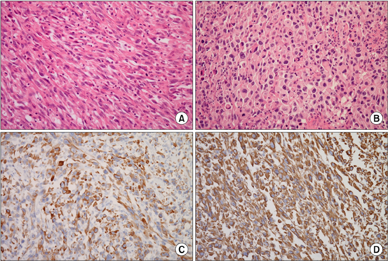
Fig. 2 Microscopically, tumor is composed of atypical spindle (A: H&E, ×200) and discohesive giant polygonal cells (B: H&E, ×200) in inflammatory background. Immunohistochemical staining, tumor cells are positive for cytokeratin (C: cytokeratin, ×200) and vimentin (D: vimentin, ×200).
Reference
-
1. Reid-Nicholson M, Idrees M, Perino G, Hytiroglou P. Sarcomatoid carcinoma of the small intestine: a case report and review of the literature. Arch Pathol Lab Med. 2004. 128:918–921.2. Kim MJ, Yu E, Ro JY. Sarcomatoid carcinoma of the gallbladder with a rhabdoid tumor component. Arch Pathol Lab Med. 2003. 127:e406–e408.3. Khan AR. Sarcomatoid carcinoma of the stomach with heterologous elements. Ann Saudi Med. 1999. 19:135–136.4. Raza MA, Mazzara PF. Sarcomatoid carcinoma of esophagus. Arch Pathol Lab Med. 2011. 135:945–948.5. Choi YY, Jeen YM, Kim YJ. Sarcomatoid carcinoma of colon: extremely poor prognosis. J Korean Surg Soc. 2011. 80:Suppl 1. S26–S30.6. Yucel AF, Kocakusak A, Arikan S, Demirbag N, Tarlaci A, Batur S. A rare cause of acute abdomen: perforated primary sarcomatoid carcinoma of the small intestine: report of a case, with a brief review of the literature. J Cancer Res Ther. 2011. 7:348–350.7. Moriwaki Y, Sugiyama M. Severe anemia inducing preshock caused by sarcomatoid carcinoma of the small intestine. Int Surg. 2009. 94:164–170.8. Dikman SH, Toker C. Enteroblastoma complicating regional enteritis. Gastroenterology. 1973. 65:462–466.9. Paik HJ, Choi YM. A case of carcinosarcoma in duodenum. J Korean Surg Soc. 1991. 41:549–553.10. Lam KY, Leung CY, Ho JW. Sarcomatoid carcinoma of the small intestine. Aust N Z J Surg. 1996. 66:636–639.
- Full Text Links
-
- Actions
-
Cited
- CITED
-
- Close
- Share
-
- Similar articles
-
- Sarcomatoid Carcinoma of the Small Intestine: A Case Report and Review of the Literature
- Sarcomatoid Carcinoma of the Duodenum: A case report
- A Surgically Resected Large Sarcomatoid Carcinoma of the Jejunum: A Case Report and Literature Review
- Sarcomatoid Renal Cell Carcinoma; Special Reference to its Distinction from Carcinosarcoma
- A Case of Sarcomatoid Cholangiocarcinoma Which Developed at the Site Previously Treated by Transarterial Chemoembolization
