Two cases of mucinous adenocarcinoma of the stomach mistaken as submucosal tumor
- Affiliations
-
- 1Department of Surgery, Gachon University, Incheon, Korea. lwk@gilhospital.com
- KMID: 2145000
- DOI: http://doi.org/10.4174/jkss.2013.84.2.118
Abstract
- A gastric carcinoma with the endoscopic features resembling submucosal tumor (SMT) is rare, and reportedly account for only 0.1% to 0.63% of all resected gastric carcinomas. The preoperative diagnosis of SMT-like gastric carcinoma is challenging, and thus, diagnosis is usually made intraoperatively or postoperatively. Furthermore, mucinous adenocarcinoma is an uncommon histologic subtype of gastric carcinoma characterized as an elevated lesion resembling SMT due to abundant mucin pools in submucosa. Here, we report two cases in which a gastric mucinous adenocarcinoma was mistaken as a SMT.
Keyword
MeSH Terms
Figure
-

Fig. 1 (A) Endoscopy revealed two large round, protruding masses in the antrum without a mucosal abnormality. (B) Endoscopic ultrasonography showed two large cystic lesions containing nonhomogenous mixed echoic masses in the proper muscle layer, without nearby lymph node enlargement. (C) Computed tomography revealed a -3.1 cm sized multilocular cystic mass in the submucosal portion of the pylorus.
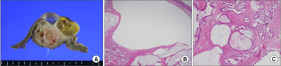
Fig. 2 (A) Cross section showing tumor infiltration into proper muscle and subserosa with normally appearing overlying mucosa. (B) The tumor was located under the intact mucosa (H&E, ×12.5). (C) The tumor showing focal papillary growth and extracelluar pools of mucin (H&E, ×100).
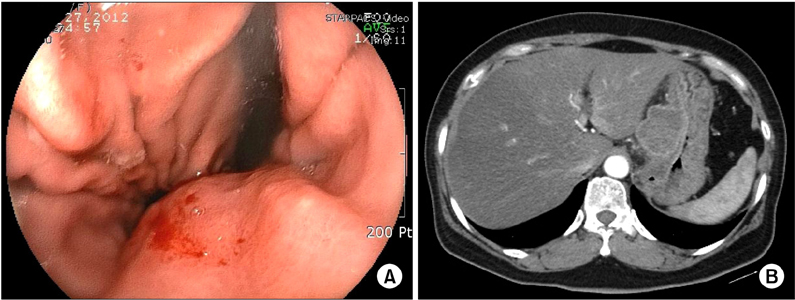
Fig. 3 (A) Endoscopy revealed an extrinsic mass effect in the lesser curvature of the midbody, and there was redness of mucosa around the lesion. However there was no evident erosion. (B) Computed tomography revealed a -5 cm sized mass with partial necrotic change in the lesser curvature side of the stomach body and multiple lymphadenopathy.
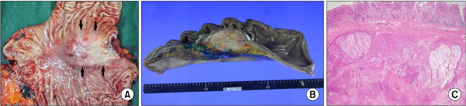
Fig. 4 (A) The mucosal surface is slightly red in comparison with the surrounding gastric mucosa but no erosion is evident. (B) On cross section, the tumor was found to have infiltrated subserosa. The tumor had a brownish tan color and necrotic change was observed in its center. (C) Low power histologic examination reveals most of the cancer cells are covered with noncancerous epithelium (H&E, ×12.5).
Cited by 1 articles
-
점액성 위선암과 비점액성 위선암 및 위반지세포암종의 임상적 비교
Honggeun Ahn, Woo Chul Chung, Yeon-Ji Kim, Seongyul Ryu, Eunsun Lim
Korean J Gastroenterol. 2020;76(6):297-303. doi: 10.4166/kjg.2020.098.
Reference
-
1. Umehara Y, Kimura T, Okubo T, Sano Y, Nakai K, Oi S, et al. Gastric carcinoma resembling submucosal tumor. Gastric Cancer. 1999. 2:191–193.2. Toyohiko Y, Tadasu S, Kazuhiko I, Shigeharu S, Satoshi S, Dai H, et al. Clinicopathological and imaging features of gastric carcinoma resembling submucosal tumor. Stomach Intest. 2003. 38:1527–1536.3. Noriya U, Hiroyasy I, Shingo I, Hiroshi S, Shuji O, Tsutomo K, et al. Mucinous adenocarcinoma of the stomach mimicking submucosal tumor: report of a case. Stomach Intest. 2003. 38:1557–1561.4. Nomura H, Mai M. Enhanced expression of CA19-9 in mucinous gastric carcinoma. Am J Gastroenterol. 1997. 92:2331–2332.5. Teraishi F, Uno F, Kagawa S, Fujiwara T, Gouchi A, Tanaka N. Advanced gastric adenocarcinoma mimicking a submucosal tumor. Endoscopy. 2007. 39:Suppl 1. E191–E192.6. Ohara N, Tominaga O, Uchiyama M, Nakano H. A case of advanced gastric cancer resembling submucosal tumor of the stomach. Jpn J Clin Oncol. 1997. 27:423–426.7. Joensuu H, Fletcher C, Dimitrijevic S, Silberman S, Roberts P, Demetri G. Management of malignant gastrointestinal stromal tumours. Lancet Oncol. 2002. 3:655–664.
- Full Text Links
-
- Actions
-
Cited
- CITED
-
- Close
- Share
-
- Similar articles
-
- A Case of Mucinous Gastric Adenocarcinoma as Submucosal Tumor
- A Patient with Jejunal Mucinous Adenocarcinoma Metastatic to the Stomach Presenting with Submucosal Tumors in the Stomach and Jejunum
- A Case of Mucinous Gastric Adenocarcinoma Mimicking Submucosal Tumor
- Collision of Adenocarcinoma and Schwannoma of the Stomach: A Case Report
- Composite Neuroendocrine Carcinoma with Adenocarcinoma of the Stomach Misdiagnosed as a Giant Submucosal Tumor
