Composite Neuroendocrine Carcinoma with Adenocarcinoma of the Stomach Misdiagnosed as a Giant Submucosal Tumor
- Affiliations
-
- 1Department of Surgery, Catholic University of Daegu School of Medicine, Daegu, Korea. hdchae@cu.ac.kr
Abstract
- A composite glandular/exocrine-endocrine carcinoma of the gastrointestinal tract is characterized by the co-existence of two adjacent, but histologically-distinct tumors in an organ. Composite glandular/exocrine-endocrine carcinomas are a special type of tumor comprised of common adenocarcinomas and neuroendocrine components that account for at least one-third of the entire tumor area. Composite tumors have been reported in a range of organs, but are relatively rare in the stomach. We report a case of a composite neuroendocrine carcinoma with an adenocarcinoma of the stomach (mixed exocrine-endocrine carcinoma), which was misdiagnosed as a giant submucosal tumor preoperatively based on esophagogastroduodenoscopy and a contrast-enhanced axial computed tomographic scan.
Keyword
MeSH Terms
Figure
-
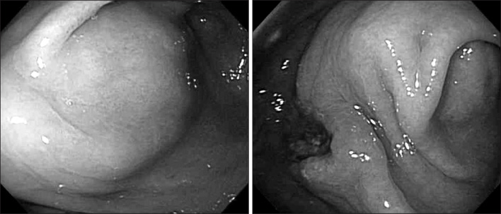
Fig. 1 Gastroduodenoscopy shows a huge, well-defined fungating submucosal mass, measuring 10 cm in its largest dimension with eccentric ulceration and an ill-defined margin in the gastric antrum, lesser curvature.
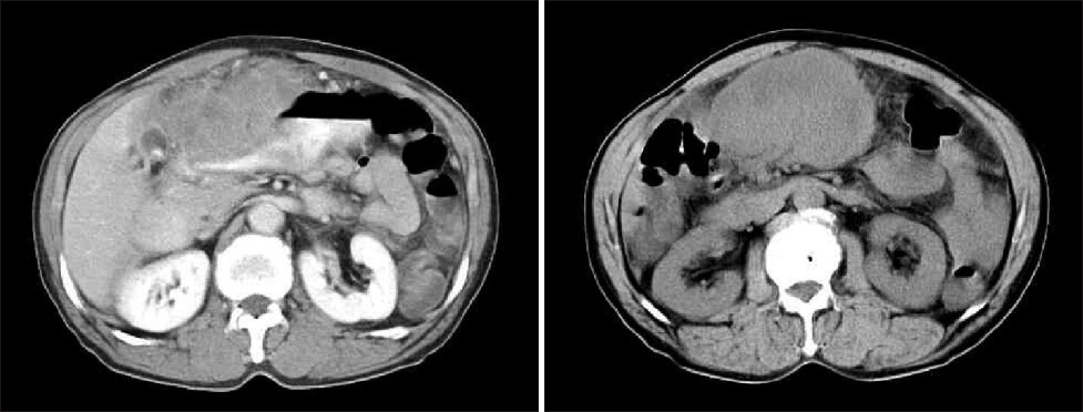
Fig. 2 Preoperative contrast-enhanced axial CT scan shows a huge, solid, ovoid, heterogenous and exophytic mass measuring approximately 15×10 cm in the prepyloric antrum of the stomach.
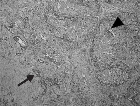
Fig. 3 The tumor is composed of conventional adenocarcinoma with anaplastic glandular nests (arrow) and neuroendocrine carcinoma with central necrosis (arrow head) (H&E, ×100).

Fig. 4 Neuroendocrine carcinoma components show strong immunopositivity for chromogranin A (left upper), but negative immunostaining of adenocarcinoma component (right lower) (Immunohistochemical stain, ×200).
Reference
-
1. Liu SW, Chen GH, Hsieh PP. Collision tumor of the stomach: a case report of mixed gastrointestinal stromal tumor and adenocarcinoma. J Clin Gastroenterol. 2002. 35:332–334.2. Fukui H, Takada M, Chiba T, Kashiwagi R, Sakane M, Tabata F, et al. Concurrent occurrence of gastric adenocarcinoma and duodenal neuroendocrine cell carcinoma: a composite tumour or collision tumours? Gut. 2001. 48:853–856.
Article3. Chejfec G, Falkmer S, Askensten U, Grimelius L, Gould VE. Neuroendocrine tumors of the gastrointestinal tract. Pathol Res Pract. 1988. 183:143–154.
Article4. Granberg D, Wilander E, Stridsberg M, Granerus G, Skogseid B, Oberg K. Clinical symptoms, hormone profiles, treatment, and prognosis in patients with gastric carcinoids. Gut. 1998. 43:223–228.
Article5. Hedenbro JL, Ekelund M, Wetterberg P. Endoscopic diagnosis of submucosal gastric lesions. The results after routine endoscopy. Surg Endosc. 1991. 5:20–23.6. Lewin K. Carcinoid tumors and the mixed (composite) glandular-endocrine cell carcinomas. Am J Surg Pathol. 1987. 11:71–86.
Article7. Fujiyoshi Y, Kuhara H, Eimoto T. Composite glandular-endocrine cell carcinoma of the stomach. Report of two cases with goblet cell carcinoid component. Pathol Res Pract. 2005. 200:823–829.
Article8. Fossmark R, Zhao CM, Martinsen TC, Kawase S, Chen D, Waldum HL. Dedifferentiation of enterochromaffin-like cells in gastric cancer of hypergastrinemic cotton rats. APMIS. 2005. 113:436–449.
Article9. Waldum HL, Rørvik H, Falkmer S, Kawase S. Neuroendocrine (ECL cell) differentiation of spontaneous gastric carcinomas of cotton rats (Sigmodon hispidus). Lab Anim Sci. 1999. 49:241–247.10. Lee EJ, Park SM, Maeng L, Lee A, Kim KM. Composite glandular-endocrine cell carcinomas of the stomach: clinicopathologic and methylation study. APMIS. 2005. 113:569–576.
Article11. Shibuya H, Azumi N, Abe F. Gastric small-cell undifferentiated carcinoma with adeno and squamous cell carcinoma components. Acta Pathol Jpn. 1985. 35:473–480.
Article12. Kim KM, Kim MJ, Cho BK, Choi SW, Rhyu MG. Genetic evidence for the multi-step progression of mixed glandular-neuroendocrine gastric carcinomas. Virchows Arch. 2002. 440:85–93.
Article13. Volante M, Rindi G, Papotti M. The grey zone between pure (neuro)endocrine and non-(neuro)endocrine tumours: a comment on concepts and classification of mixed exocrine-endocrine neoplasms. Virchows Arch. 2006. 449:499–506.
Article14. Elias D, Cavalcanti de, Eggenspieler P, Plaud B, Ducreux M, Spielmann M, et al. Resection of liver metastases from a noncolorectal primary: indications and results based on 147 monocentric patients. J Am Coll Surg. 1998. 187:487–493.
Article15. Park BS, Jo TY, Seo HI, Kim HS, Kim DH, Jeon TY, et al. Gastric collision tumor (adenocarcinoma and neuroendocrine carcinoma) diagnosed as a advanced gastric cancer. J Korean Surg Soc. 2007. 73:173–177.
- Full Text Links
-
- Actions
-
Cited
- CITED
-
- Close
- Share
-
- Similar articles
-
- A Gastric Composite Tumor with an Adenocarcinoma and a Neuroendocrine Carcinoma: A Case Report
- A Case of a Gastric Composite Tumor with an Adenocarcinoma and a Large Cell Neuroendocrine Carcinoma
- Gastric Collision Tumor (Adenocarcinoma and Neuro-endocrine Carcinoma): A Report of Two Cases
- Composite Carcinoma-Carcinoid Tumor of Stomach: Report of a case
- Gastric Adenocarcinoma with Coexistent Hepatoid Adenocarcinoma and Neuroendocrine Carcinoma: A Case Report
