J Korean Soc Magn Reson Med.
2013 Dec;17(4):308-311. 10.13104/jksmrm.2013.17.4.308.
Deep Sylvian Meningioma in a 43-Year-Old Man: A Case Report
- Affiliations
-
- 1Department of Radiology, Keimyung University, Dongsan Medical Center, Daegu, Korea. hyukwonchang@korea.com
- 2Department of Pathology, Keimyung University, Dongsan Medical Center, Daegu, Korea.
- 3Department of Neurosurgery, Keimyung University, Dongsan Medical Center, Daegu, Korea.
- KMID: 2144332
- DOI: http://doi.org/10.13104/jksmrm.2013.17.4.308
Abstract
- Deep sylvian meningioma is a rare form of meningiomas. So far, only 4 cases including the present one have been reported in South Korea. A 43-year-old man without any previous medical history presented to our hospital with seizure. There was a rim enhancing mass in the right deep sylvian fissure without dural attachment on magnetic resonance images. Surgical resection of the mass revealed the lesion to be a meningioma in this patient.
Keyword
Figure
-
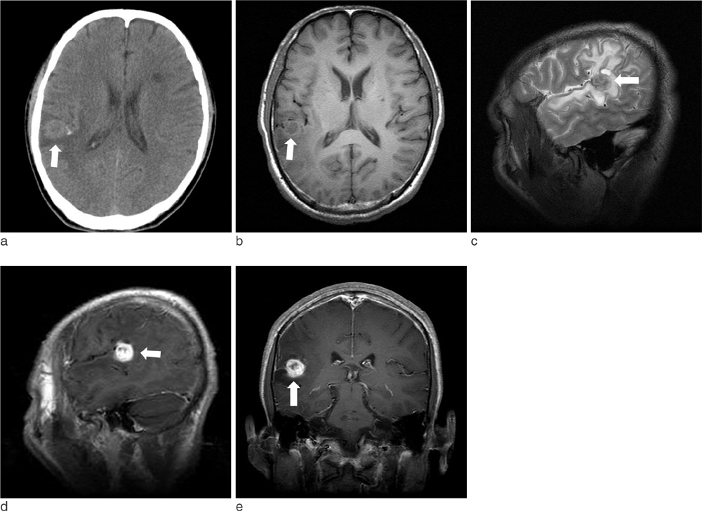
Fig. 1 The initial CT (a) study revealed a 15×16×16 mm rim enhancing mass located in the distal portion of right sylvian fissure with minimal calicifications. On MRI study, this lesion was found to have iso-signal intensity in T1 (b) and T2 (c) weighted images with surrounding adjacent edema. In T1 contrast phase (d, e), the mass showed ring like enhancement.
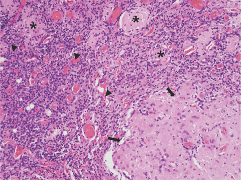
Fig. 2 The mass showing meningothelial proliferative lesion (arrows) and surrounding lymphoplasmacytic infiltrative background (arrowheads). Scattered meningioma component are noted (asterixs)
Reference
-
1. Zhang J, Chi LY, Meng B, Li F, Zhu SG. Meningioma without dural attachment: case report, classification, and review of the literature. Surg Neurol. 2007; 67:535–539.2. Cushing H, Eisenhardt L. Meningiomas. Their Classification, Regional Behaviour, Life History, and Surgical End Results. New York: Hafner Publishing Company;1969. p. 133–168.3. Cecchi PC, Campello M, Rizzo P, Mair K, Schwarz A. Atypical meningioma of the sylvian fissure. J Clin Neurosci. 2009; 16:1234–1239.4. Cho BK, Wang KC, Chang KH, Chi JG. Deep sylvian meningioma in a child. Childs Nerv Syst. 1990; 6:228–230.5. Moon BJ, Choi JY, Park YG, Chung SS. Deep sylvian meningioma: case report. J Korean Neurosurg Soc. 2003; 33:218–221.6. Chae MP, Song SW, Park SH, Park CK. Experience with 5-aminolevulinic acid in fluorescence-guided resection of a deep sylvian meningioma. J Korean Neurosurg Soc. 2012; 52:558–560.7. Jung YS, Song YJ. Meningioma in a 20-month-old boy. J Korean Neurosurg Soc. 2012; 51:219–221.8. Choi YM, Kim TY, Kim JM. A case of meningioma in temporo-occipital lobe without dural attachment in a 14-yer-old girl: case report. J Korean Neurosurg Soc. 1996; 25:1101–1107.9. Ko BS, Jung S, Jung TY, Kim IY. Intraparenchymal sylvian fissure meningioma. J Korean Neurosurg Soc. 2007; 41:120–122.10. Aras Y, Akcakaya MO, Aydoseli A, Izgi N. Staged surgery for sylvian fissure meningiomas without dural attachment: report of two cases. Clin Neurol Neurosurg. 2013; 115:1527–1529.
- Full Text Links
-
- Actions
-
Cited
- CITED
-
- Close
- Share
-
- Similar articles
-
- Deep Sylvian Meningioma: Case Report
- Experience with 5-Aminolevulinic Acid in Fluorescence-Guided Resection of a Deep Sylvian Meningioma
- A Subcortical Anaplastic Meningioma
- Cranial meningioma of unusual location: two cases report
- Intraventricular Hemorrhage Caused by Lateral Ventricular Meningioma: A Case Report
