Experience with 5-Aminolevulinic Acid in Fluorescence-Guided Resection of a Deep Sylvian Meningioma
- Affiliations
-
- 1University of Melbourne, Melbourne, Victoria, Australia.
- 2Department of Neurosurgery, Seoul National University College of Medicine, Seoul National University Hospital, Seoul, Korea. nsckpark@snu.ac.kr
- 3Department of Pathology, Seoul National University College of Medicine, Seoul National University Hospital, Seoul, Korea.
- KMID: 1426268
- DOI: http://doi.org/10.3340/jkns.2012.52.6.558
Abstract
- The 5-aminolevulinic acid (5-ALA)-induced tumor fluorescence is a useful intraoperative marker for the diagnosis and the detection of various malignancies, but its use in meningioma is only reported infrequently. In meningioma, a complete resection of the tumor mass is crucial for the prevention of recurrence and postoperative morbidities. Deep sylvian meningioma is a rare type of meningioma where complete tumor removal is complicated by its deep anatomical location and close involvement with the middle cerebral artery. From our experience, 5-ALA-mediated fluorescence facilitated a safe excision whilst preserving critical neurovascular structures. To our best knowledge, this is first report from use of 5-ALA in a deep sylvian meningioma.
Keyword
Figure
-

Fig. 1 A preoperative non-contrast enhanced axial CT (A) shows an area of high density in the left temporal lobe, most likely a calcification. An axial T2-weighted magnetic resonance (MR) image without contrast (B) shows a mass lesion of mixed signal intensity with edema located deep in the left Sylvian fissure. It displays heterogeneous contrast enhancement on a T1-weighted MR image (C). A post-operative axial T2-weighted MR image taken within 48 hours of the surgery (D) shows the resection cavity with a residual tumor tissue strongly adherent to the middle cerebral artery.
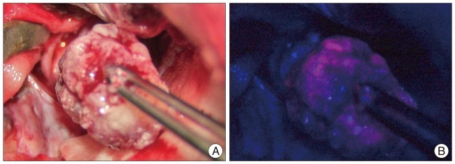
Fig. 2 An intra-operative photograph of the excised mass shone under the white light (A) is taken using the surgical microscope. Under the violet-blue excitation light (B), it exhibits strong red fluorescence, confirming the presence of a neoplasm.

Fig. 3 Pathological examination confirms the diagnosis of the fluorescent mass as a meningioma. Microscopically, the tissue demonstrates syncytial growth of meningothelial cells with bland looking nuclei and psammoma bodies (H&E stain; original magnification ×200).
Cited by 1 articles
-
Experience Profiling of Fluorescence-Guided Surgery II: Non-Glioma Pathologies
So Young Ji, Jin Wook Kim, Chul-Kee Park
Brain Tumor Res Treat. 2019;7(2):105-111. doi: 10.14791/btrt.2019.7.e39.
Reference
-
1. Arai T, Tani S, Isoshima A, Nagashima H, Joki T, Takahashi-Fujigasaki J, et al. [Intraoperative photodynamic diagnosis for spinal ependymoma using 5-aminolevulinic acid : technical note]. No Shinkei Geka. 2006; 34:811–817. PMID: 16910494.2. Cecchi PC, Campello M, Rizzo P, Mair K, Schwarz A. Atypical meningioma of the sylvian fissure. J Clin Neurosci. 2009; 16:1234–1239. PMID: 19497747.
Article3. Chiocca EA, Boviatsis EJ, Westmark RM, Short MP, Richardson EP, Zervas NT. Deep sylvian fissure meningioma without dural attachment in an adult : case report. Neurosurgery. 1994; 35:944–946. discussion 946. PMID: 7838346.4. Coluccia D, Fandino J, Fujioka M, Cordovi S, Muroi C, Landolt H. Intraoperative 5-aminolevulinic-acid-induced fluorescence in meningiomas. Acta Neurochir (Wien). 2010; 152:1711–1719. PMID: 20535506.
Article5. Grant WE, Hopper C, MacRobert AJ, Speight PM, Bown SG. Photodynamic therapy of oral cancer : photosensitisation with systemic aminolaevulinic acid. Lancet. 1993; 342:147–148. PMID: 7687318.
Article6. Gupta AK, Ryder JE. Photodynamic therapy and topical aminolevulinic acid : an overview. Am J Clin Dermatol. 2003; 4:699–708. PMID: 14507231.7. Kajimoto Y, Kuroiwa T, Miyatake S, Ichioka T, Miyashita M, Tanaka H, et al. Use of 5-aminolevulinic acid in fluorescence-guided resection of meningioma with high risk of recurrence. Case report. J Neurosurg. 2007; 106:1070–1074. PMID: 17564181.
Article8. Ma L, Xiao SY, Zhang YK. Atypical meningioma of sylvian fissure with a 20-year history : a rare case report. Neurol Sci. 2012; 33:143–145. PMID: 21617950.
Article9. Miyahara K, Ichikawa T, Yagishita S, Mukaihara S, Okada T, Kaku S, et al. [Deep sylvian meningioma without dural attachment : a case report]. No Shinkei Geka. 2011; 39:1067–1072. PMID: 22036818.10. Morofuji Y, Matsuo T, Hayashi Y, Suyama K, Nagata I. Usefulness of intraoperative photodynamic diagnosis using 5-aminolevulinic acid for meningiomas with cranial invasion : technical case report. Neurosurgery. 2008; 62:102–103. discussion 103-104. PMID: 18424972.11. Ruge JR, Liu J. Use of 5-aminolevulinic acid for visualization and resection of a benign pediatric brain tumor. J Neurosurg Pediatr. 2009; 4:484–486. PMID: 19877785.
Article12. Samson Sujit Kumar G, Rajshekhar V. Deep sylvian meningioma : a case report and review of literature. Childs Nerv Syst. 2009; 25:129–132. PMID: 18712398.
Article13. Stummer W, Pichlmeier U, Meinel T, Wiestler OD, Zanella F, Reulen HJ. Fluorescence-guided surgery with 5-aminolevulinic acid for resection of malignant glioma : a randomised controlled multicentre phase III trial. Lancet Oncol. 2006; 7:392–401. PMID: 16648043.
Article
- Full Text Links
-
- Actions
-
Cited
- CITED
-
- Close
- Share
-
- Similar articles
-
- Fluorescence Guided Surgery with 5-Aminolevulinic Acid for Resection of Spinal Cord Ependymomas
- 5-Aminolevulinic Acid Fluorescence Discriminates the Histological Grade of Extraventricular Neurocytoma
- Deep Sylvian Meningioma: Case Report
- Deep Sylvian Meningioma in a 43-Year-Old Man: A Case Report
- A reliable method for the adjustment of urinary delta-aminolevulinic acid concentration
