The retrospective study of marginal bone loss around dental implants according to different autogenous bone grafts
- Affiliations
-
- 1Department of Implantology, Graduate School of Clinical Dentistry, Ewha Womans University, Seoul, Korea.sjsj7777@ewha.ac.kr
- 2Department of Oral and Maxillofacial Surgery, School of Medicine, Ewha Womans University, Seoul, Korea.
- KMID: 2136985
- DOI: http://doi.org/10.5125/jkaoms.2011.37.6.483
Abstract
- INTRODUCTION
This study examined the cumulative resorption of implants placed in a severely atrophic mandible and analyzed the radiologic bone resorption in the marginal bone, after an autogenous bone graft including both block and particulates that had been harvested from the ramus and iliac crest.
MATERIALS AND METHODS
A retrospective study was performed on patients who had bone grafts for augmentation followed by implant installation in the mandible area from 2003 to 2008. Twelve patients (6 men and 6 women) who received 34 implants in the augmented sites were evaluated. Cumulative radiologic resorption around the implants was measured immediately, 3 months, 6 months and 12 months after implant installation surgery.
RESULTS
The installed implant in grafted bone showed 0.84 mm marginal bone resorption after 3 months and 50% total cumulative resorption after 1 year. The mean marginal bone resorption around the implant installed in the grafted bone was 0.44 mm after 3 months, 0.52 mm after 1 year, after which it stabilized. The implant survival rate was 97% (failed implant was 1/34). Marginal bone resorption of the installed implant in the autogenous onlay block bone grafts was 0.98 mm after 3 months, which was significantly higher than that of a particulated bone graft (0.74 mm) (P<0.05).
CONCLUSION
An autogenous graft including block type and particulate type is a predictable procedure for the use of dental implants in a severely atrophic mandible. Implant placement in augmented areas show a relatively high survival and minimal bone loss, as revealed by a radiologic evaluation.
Keyword
MeSH Terms
Figure
-
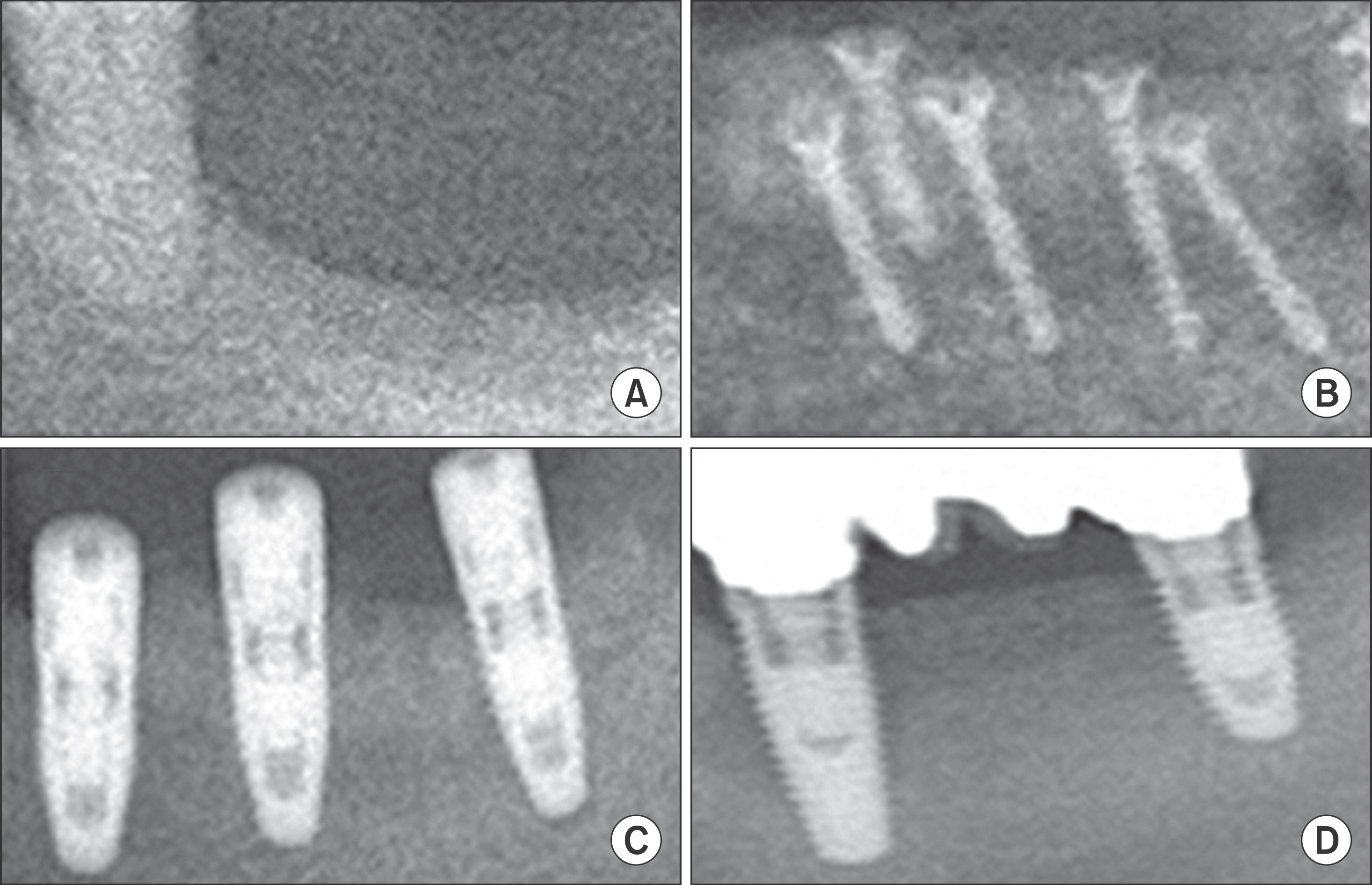
Fig. 1. Autogenous block bone graft, implant installation and functional loading. These figures were collected from different patients who underwent ramal bone graft and implant installation surgery. A. Pre-operative radiography. B. Immediate post-operative radiography after autogenous block bone graft operation. C. Immediate post-operative radiography after implant fixtures installation operation. D. Follow-up radiography after functional loading. Tae-Yi Kim et al: The retrospective study of marginal bone loss around dental implants according to different autogenous bone grafts. J Korean Assoc Oral Maxillofac Surg 2011

Fig. 2. Simultaneous implant installation with autogenous particulated bone and xenogenic bone graft. These figures were collected from different patients who underwent particulated autogenous bone and xenogenic bone graft and implant installation surgery. A. Pre-operative radiography. These previous implants were removed at bone graft surgery because of those mobility and pain. B. Immediate postoperative radiography after mixed particulated bone graft and implant installation. C. Follow-up post-operative radiography after functional loading. Tae-Yi Kim et al: The retrospective study of marginal bone loss around dental implants according to different autogenous bone grafts. J Korean Assoc Oral Maxillofac Surg 2011
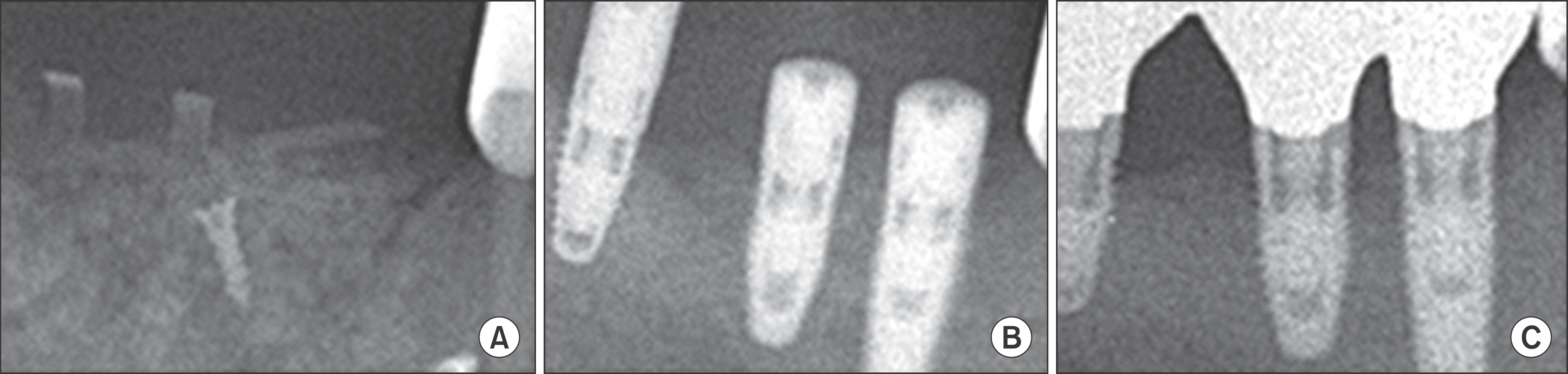
Fig. 3. Delayed implant installation after guided bone regeneration technique with autogenous particulated bone. A. Immediate postoperative radiography after autogenous particulated bone graft. B. Immediate post-operative radiography after 7-month delayed implant installation. C. Follow-up radiography after implant functional loading. Tae-Yi Kim et al: The retrospective study of marginal bone loss around dental implants according to different autogenous bone grafts. J Korean Assoc Oral Maxillofac Surg 2011
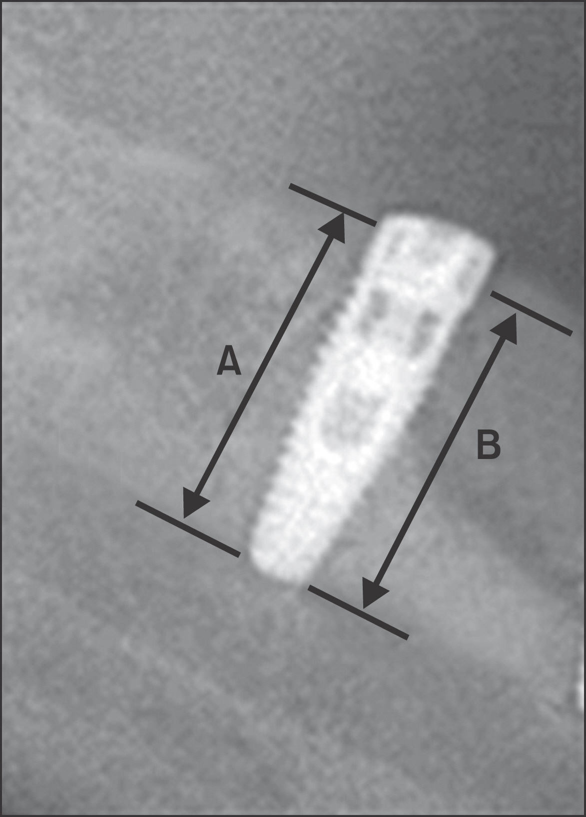
Fig. 4. Formula for calculation of bone level and height. Average real length was calculated from (A+B/2 ×magnificent rate). Tae-Yi Kim et al: The retrospective study of marginal bone loss around dental implants according to different autogenous bone grafts. J Korean Assoc Oral Maxillofac Surg 2011
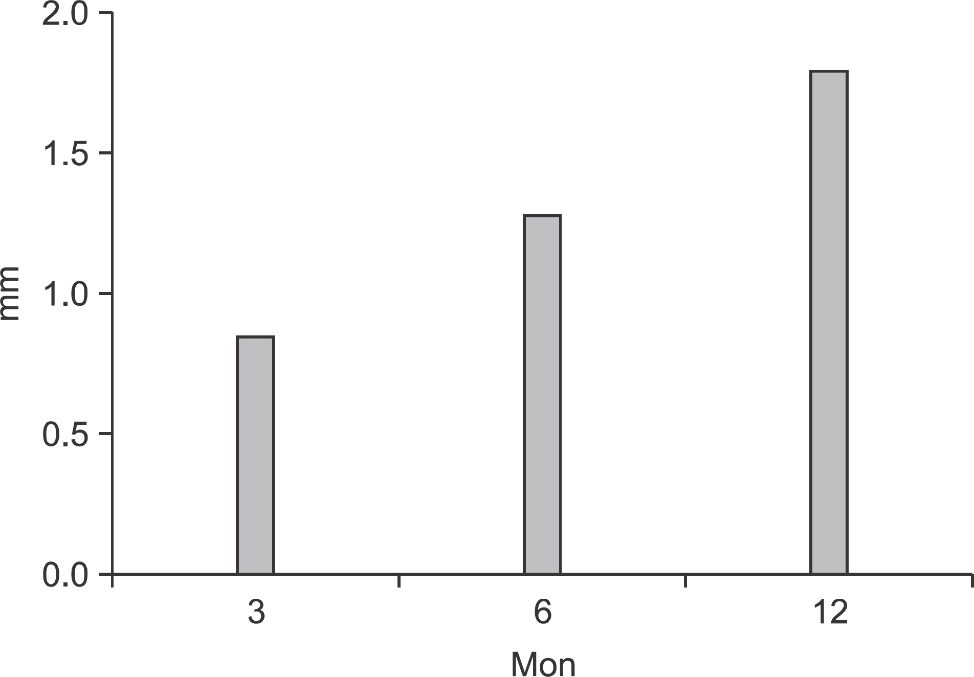
Fig. 5. Cumulative marginal bone resorption after implant placement in/with the grafted bone. Tae-Yi Kim et al: The retrospective study of marginal bone loss around dental implants according to different autogenous bone grafts. J Korean Assoc Oral Maxillofac Surg 2011
- Full Text Links
-
- Actions
-
Cited
- CITED
-
- Close
- Share
-
- Similar articles
-
- Clinical usages of ramal autogenous bone grafts in dental implant surgery
- Retrospective study on marginal bone loss around maxillary anterior implants with or without bone graft
- Influence of crown-to-implant ratio on periimplant marginal bone loss in the posterior region: a five-year retrospective study
- Comparison of marginal bone loss of dental implants and adjacent teeth in the same interproximal unit: a retrospective study with follow-up over 2 years after prosthesis delivery
- Marginal bone level changes in association with different vertical implant positions: a 3-year retrospective study
