Clinical and scanning electron microscopic analysis of fractured dental implants: a retrospective clinical analysis
- Affiliations
-
- 1Department of Oral and Maxillofacial Surgery, School of Dentistry, Wonkwang University, Iksan, Korea. kkhoms@daum.net
- 2Wonkwang Dental Research Institute, Iksan, Korea.
- KMID: 2136975
- DOI: http://doi.org/10.5125/jkaoms.2012.38.6.371
Abstract
- Many longitudinal studies have reported the successful osseointegration of dental implants, with survival rates approaching 90-95%. However, implants regarded as a "success" may have also failed to undergo osseointegration. A variety of complications and failures have been observed, including implant fracture - a rare and delayed biomechanical complication with serious clinical outcomes. Given the increasing popularity of dental implants, an increase in the number of failures due to late fractures is expected. This study sought to determine the rate of implant fractures and factors associated with its development. This retrospective evaluation analyzed implants placed at Wonkwang Dental Hospital (from 1996 to the present). In our study we found that the frequency of dental implant fractures was very low (0.23%, 8 implant fractures out of 3,500 implants placed). All observed fractures were associated with hybrid-surface threaded implants (with diameter of 4.0 or 3.75 mm). Prosthetic or abutment screw loosening preceded implant fracture in a majority of these cases.
MeSH Terms
Figure
-
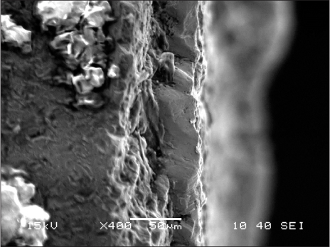
Fig. 1 Scanning electron microscopy views of the fracture. High power view of the fractured surface of titanium implant; magnification ×400. Fracture cross-section has a different plan.
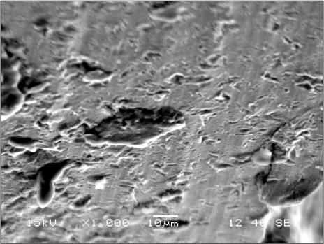
Fig. 2 It was possible to see that the titanium surface presented many porosities.
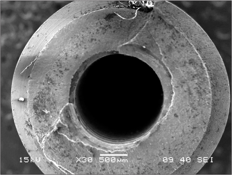
Fig. 3 At low-power scanning electron microscopic magnification (original magnification ×30), it is possible to observe that no porosity is present inside the titanium and that the fractures appeared in different planes. The fractured surface exhibits a dimpled aspect, characteristic of a tensile fracture.
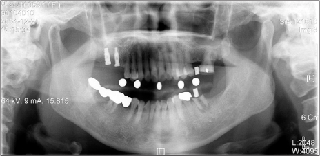
Fig. 4 Initial panoramic radiograph. This view shows missing state (#26, #36, #37) where implant is placed.
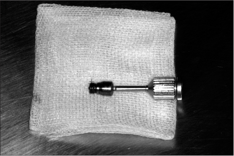
Fig. 5 Removal finding of healing abutment and coronal fragment of implant.

Fig. 6 Final prosthodontic view finding (Patient 1).
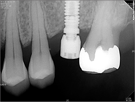
Fig. 7 At 3-year follow-up radiograph after final prosthodontic restoration, implant fixture fracture was evident.
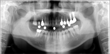
Fig. 8 The panoramic radiograph of a 50 year old male with existing restoration were 2 ITI implant system on left maxillary posterior molar area. These had been extracted 1 month ago (2006 Jan 20) previously possibly due to endodontic or structural problem.
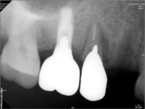
Fig. 9 Upper second premolar was demonstrated mobility and fractured at cervical area. Overload-induced bone resorption seemed to precede implant fracture (Patient 5). There was no fracture sign in implant fixture.
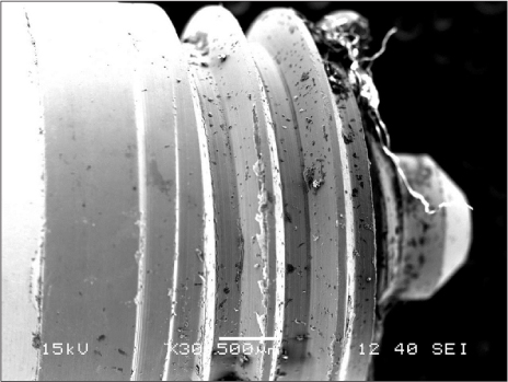
Fig. 10 At low-power scanning electron microscopic magnification (×30), tearing striation are present (Case No. 5).
Reference
-
1. Buser D, Mericske-Stern R, Bernard JP, Behneke A, Behneke N, Hirt HP, et al. Long-term evaluation of non-submerged ITI implants. Part 1: 8-year life table analysis of a prospective multi-center study with 2359 implants. Clin Oral Implants Res. 1997. 8:161–172.
Article2. Adell R, Lekholm U, Rockler B, Brånemark PI. A 15-year study of osseointegrated implants in the treatment of the edentulous jaw. Int J Oral Surg. 1981. 10:387–416.
Article3. Tolman DE, Laney WR. Tissue-integrated prosthesis complications. Int J Oral Maxillofac Implants. 1992. 7:477–484.
Article4. Balshi TJ. An analysis and management of fractured implants: a clinical report. Int J Oral Maxillofac Implants. 1996. 11:660–666.5. Rangert B, Krogh PH, Langer B, Van Roekel N. Bending overload and implant fracture: a retrospective clinical analysis. Int J Oral Maxillofac Implants. 1995. 10:326–334.6. Muroff FI. Removal and replacement of a fractured dental implant: case report. Implant Dent. 2003. 12:206–210.
Article7. Gargallo Albiol J, Satorres-Nieto M, Puyuelo Capablo JL, Sánchez Garcés MA, Pi Urgell J, Gay Escoda C. Endosseous dental implant fractures: an analysis of 21 cases. Med Oral Patol Oral Cir Bucal. 2008. 13:E124–E128.8. Morgan MJ, James DF, Pilliar RM. Fractures of the fixture component of an osseointegrated implant. Int J Oral Maxillofac Implants. 1993. 8:409–414.9. Piattelli A, Piattelli M, Scarano A, Montesani L. Light and scanning electron microscopic report of four fractured implants. Int J Oral Maxillofac Implants. 1998. 13:561–564.10. Zetterqvist L, Feldman S, Rotter B, Vincenzi G, Wennström JL, Chierico A, et al. A prospective, multicenter, randomized-controlled 5-year study of hybrid and fully etched implants for the incidence of peri-implantitis. J Periodontol. 2010. 81:493–501.
Article11. Eckert SE, Meraw SJ, Cal E, Ow RK. Analysis of incidence and associated factors with fractured implants: a retrospective study. Int J Oral Maxillofac Implants. 2000. 15:662–667.12. Jemt T, Lekholm U. Oral implant treatment in posterior partially edentulous jaws: a 5-year follow-up report. Int J Oral Maxillofac Implants. 1993. 8:635–640.13. Schwarz MS. Mechanical complications of dental implants. Clin Oral Implants Res. 2000. 11:Suppl 1. 156–158.
Article14. Levine RA, Clem DS 3rd, Wilson TG Jr, Higginbottom F, Saunders SL. A multicenter retrospective analysis of the ITI implant system used for single-tooth replacements: preliminary results at 6 or more months of loading. Int J Oral Maxillofac Implants. 1997. 12:237–242.15. Siddiqui AA, Caudill R. Proceedings of the 4th International Symposium on Implant Dentistry: Focus on Esthetics. San Diego, California, January 27-29, 1994. Abstracts. J Prosthet Dent. 1994. 72:623–634.
Article16. Turpin YL, Tardivel RD, Tallec A, Le Menn AC. Corrosion susceptibility of titanium covered by dental cements. Dent Mater. 2000. 16:57–61.
Article17. Piao CM, Lee JE, Koak JY, Kim SK, Rhyu IC, Han CH, et al. Marginal bone loss around three different implant systems: radiographic evaluation after 1 year. J Oral Rehabil. 2009. 36:748–754.
Article18. Jung YC, Han CH, Lee KW. A 1-year radiographic evaluation of marginal bone around dental implants. Int J Oral Maxillofac Implants. 1996. 11:811–818.19. Yokoyama K, Ichikawa T, Murakami H, Miyamoto Y, Asaoka K. Fracture mechanisms of retrieved titanium screw thread in dental implant. Biomaterials. 2002. 23:2459–2465.
Article
- Full Text Links
-
- Actions
-
Cited
- CITED
-
- Close
- Share
-
- Similar articles
-
- A study on fitness of several domestic implant fixture and abutment screws
- Energy-dispersive X-ray spectroscopic investigation of a fractured non-submerged dental implant associated with abutment fracture
- Implant Surface Changes Observed with Confocal and Electron Microscopy After Probing with Metal and Plastic Periodontal Probes
- Isolation and Identification of Melanosomes from Human Hair
- Electron Microprobe Analysis and Tissue Reaction around Titanium Alloy Spinal Implants
