Principles of Quality Controlled Endoscopic Submucosal Dissection with Appropriate Dissection Level and High Quality Resected Specimen
- Affiliations
-
- 1Department of Endoscopy, Kobe University Hospital, Kobe, Japan. toyonaga@med.kobe-u.ac.jp
- 2Department of Pathology, Kishiwada Tokushukai Hospital, Osaka, Japan.
- 3Frontier Medical Science in Gastroenterology, Kobe University School of Medicine, Kobe, Japan.
- 4Translational Gastroenterology Unit, John Radcliffe Hospital, Oxford, UK.
Abstract
- Endoscopic submucosal dissection (ESD) has enabled en bloc resection of early stage gastrointestinal tumors with negligible risk of lymph node metastasis, regardless of tumor size, location, and shape. However, ESD is a relatively difficult technique compared with conventional endoscopic mucosal resection, requiring a longer procedure time and potentially causing more complications. For safe and reproducible procedure of ESD, the appropriate dissection of the ramified vascular network in the level of middle submucosal layer is required to reach the avascular stratum just above the muscle layer. The horizontal approach to maintain the appropriate depth for dissection beneath the vascular network enables treatment of difficult cases with large vessels and severe fibrosis. The most important aspect of ESD is the precise evaluation of curability. This approach can also secure the quality of the resected specimen with enough depth of the submucosal layer.
Keyword
Figure
-

Fig. 1 (A-C) The worst postulated sequence of possible events of endoscopic submucosal dissection. The bleeding occured just after the beginning of incision and dissection. The procedure was finally finished with frequent hemostasis with difficulty. The edge of incision was burned to be tough, and the entry of the devices into the submucosal layer was impossible beneath the incised mucosa, like a shell closing its husk. The entry into the submucosal layer was carried out from other mucosa, however the massive bleeding occurred during the dissection.
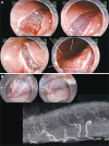
Fig. 2 The structure of vessels in the gastrointestinal luminal wall. (A) The vessel network in the submucosal layer. The shallow mucosal incision just below the muscularis mucosae. Smooth incision with less hemorrhage is enabled, and the horizontal vessel network is seen through at the middle level of the submucosal layer. (B) The large vessels in the gastrointestinal luminal wall penetrate the muscle layer vertically and then inflow horizontally at the level of middle submucosal layer forming the ramified vascular network. In the area with high density of vessels, the faciae-like layer is formed with the ramified vascular network and the perivascular fibrotic tissue, and the layer containing fewer vessels and fibrotic tissue exists below the faciae and just above the muscular propria. Smooth dissection with less hemorrhage is enabled by maintaining the appropriate depth of dissection at the layer with fewer vessels and fibrotic tissue and by coagulating the penetrating vessels before dissection.

Fig. 3 (A) An early stage gastric cancer in the posterior wall of body. (Ba) The horizontal vessel network in the submucosal layer is partially transected to reach the appropriate depth of dissection. The large vessels vertically penetrate the muscle layer and ramify horizontally at the middle level of the submucosal layer. (Bb) The ramified vessel network in the submucosal layer also connects the edges of incision. The certain dissection of this network allows the entry into the appropriate depth of submucosal dissection by broadening the groove made by the mucosal incision. (C) Once the appropriate depth of submucosal dissection has been obtained, the smooth dissection is enabled with first recognizing and properly dissecting the occasional penetrating vessels.

Fig. 4 The difference of the density of vessels. (A) The extremely characteristic muscle layers named the oblique muscle layers are symmetrically seen in the anterior/posterior regions of the gastric body. The muscle layer is circularly absent at the inlet of the large vessel. (B) The density and thickness of the vessels in the gastric antrum overwhelmingly differs from those in the gastric body. (Ba) In the antrum, the density of vessel in the submucosal layer is low, and the fibrosis is also minimal, and these allow easy mucosal incision and submucosal dissection. (Bb) In the lesser curvature of the gastric body where the oblique muscle layers exist, as the blood vessels do not diverge frequently and the density of blood vessels are low as in the gastric antrum, the procedure is rather easy if the large penetrating vessels are not hurt by mistake. (Bc) On the other hand, in the anterior/posterior walls of the gastric body, where the oblique muscle layers exist, the greater curvature of the gastric body and lower rectum, the density of blood vessels is high and the diverged vessel network is inevitably hurt, if careful attention is not paid to the depth of mucosal incision and submucosal dissection.

Fig. 5 Laterally spreading tumor: granular type in rectum. (A) Subtotal circumferential lesion. The anal side of lesion reached to the anal canal. The rectum, especially lower rectum and anal canal have largest blood vessels and also highest vessel density in the rectum. The shallow mucosal incision from the anal side was performed not to damage the vessels, and then the vessel network beneath the edges of incision was transected to reach the appropriate depth of dissection. (B) Bleeding does not occur if the dissection was performed beneath the horizontal vessel network while averting the penetrating vessels. The structure of blood vessels in the rectum was clearly recognized. The muscle layer is absent at the inlet of the large vessel in the shape of a wedge.

Fig. 6 Quality controlled endoscopic submucosal dissection (ESD). (A) The technical tip to maintain the appropriate depth of submucosal dissection (upper). A shallow mucosal incision is performed not to cause bleeding. The horizontal vessel network is disclosed by dissecting the space between the vessels that can be seen through in the submucosal layer. After precoagulation and dissection of the vessel network to scoop up from the bottom of the vessel into the lumen, the edge of mucosal incision easily expands and the transparent hood itself can get into the submucosal layer and then a mucosal flap is formed. The technical tip to maintain the appropriate depth of dissection (lower). Once the blood vessels are damaged by mistake, the field of vision is lost and subsequent procedure becomes difficult, even after the appropriate depth of dissection has been obtained. The measure is to dissect with occasional local injection beneath the vessel network while maintaining the field of vision by the transparent hood. Especially, it is always necessary to distinguish the recognized vessels whether these are horizontal small branches of the vessel network or vertical large trunks of the penetrating vessels. (B) The resected specimen obtained by quality controlled ESD. The resected specimen contains thick submucosal layer including the whole vessel network in the submucosal layer. The appropriate depth of submucosal layer has most sparse and little electric resistance and the resected specimens are minimally injured by the coagulation.

Fig. 7 Endoscopic submucosal dissection (ESD) of the lesions with severe fibrosis. (A) Laterally spreading tumor: non granular type in the colon. A mucosal flap was formed by dissection of the area with little fibrosis as much as possible after reaching the appropriate depth of submucosal dissection. The transparent hood could get into the submucosal layer despite the fibrosis because the appropriate depth of dissection has already been obtained. The fibrotic area can be safely and certainly dissected by dividing the muscle layer and lesions using the sheath of Flush knife and also connecting the lines of muscle layer at both sides. (B) The view after dissection of the fibrotic area and the resected specimen. The lesion was accompanied with severe fibrosis; however, the high-quality resected specimen was obtained with the entire vessel network. In ESD of lesion with severe fibrosis, it is important first to reach the appropriate depth of submucosal dissection (H&E stain, ×12.5).

Fig. 8 The resected specimen of a case which was revealed to be outside the criteria. 0-IIc lesion in the posterior wall of the gastric body. It was diagnosed as a mucosal cancer and endoscopic submucosal dissection was performed. The resected specimen was obtained with thick submucosal layer containing all of vessel network. The submucosal invasion to the depth of 260 µm and lymphovascular invasion was recognized (H&E stain, ×40). If the dissection in the shallow submucosal layer was performed, these important findings might not be contained in the resected specimen and remain in the artificial ulcer of the resection site. If the vessel network was damaged and repetitive hemostasis was performed, these findings could not be recognized. These low-quality resection and specimen might lead to misjudge the case as curatively resected. M, mucosal; SM, submucosal; tub., tubular adenocarcinoma; ly, lymphatic invasion; LM, lateral margin; VM, vertical margin; EC, curativity C.

Fig. 9 The resected specimen of a case which was confirmed to be within the criteria. The tumor invaded into the deep layer of the submucosa, but the muscularis mucosae covered this area without break (H&E stain, ×40). This was diagnosed as mucosal cancer. If the dissection was performed in the shallow layer of the submucosa and the muscularis mucosae was damaged, the specimen could be judged as massively invasive submucosal cancer with positive vertical margin of tumor. M, mucosal; tub., tubular adenocarcinoma; ly, lymphatic invasion; LM, lateral margin; VM, vertical margin; EA, curativity A.
Reference
-
1. Hirao M, Masuda K, Asanuma T, et al. Endoscopic resection of early gastric cancer and other tumors with local injection of hypertonic saline-epinephrine. Gastrointest Endosc. 1988; 34:264–269. PMID: 3391382.
Article2. Gotoda T, Kondo H, Ono H, et al. A new endoscopic mucosal resection procedure using an insulation-tipped electrosurgical knife for rectal flat lesions: report of two cases. Gastrointest Endosc. 1999; 50:560–563. PMID: 10502182.
Article3. Ono H, Kondo H, Gotoda T, et al. Endoscopic mucosal resection for treatment of early gastric cancer. Gut. 2001; 48:225–229. PMID: 11156645.
Article4. Oyama T, Kikuchi Y. Endoscopic mucosal resection in the upper GI tract-Hook knife EMR method. Minim Invasive Ther Allied Technol. 2002; 11:291–295.5. Yahagi N, Fujishiro M, Kakushima N, et al. Endoscopic submucosal dissection for early gastric cancer using the tip of an electrosurgical snare (thin type). Dig Endosc. 2004; 16:34–38.
Article6. Rösch T, Sarbia M, Schumacher B, et al. Attempted endoscopic en bloc resection of mucosal and submucosal tumors using insulated-tip knives: a pilot series. Endoscopy. 2004; 36:788–801. PMID: 15326574.
Article7. Neuhaus H, Costamagna G, Devière J, et al. Endoscopic submucosal dissection (ESD) of early neoplastic gastric lesions using a new double-channel endoscope (the "R-scope"). Endoscopy. 2006; 38:1016–1023. PMID: 17058167.
Article8. Kodashima S, Fujishiro M, Yahagi N, Kakushima N, Omata M. Endoscopic submucosal dissection using flexknife. J Clin Gastroenterol. 2006; 40:378–384. PMID: 16721217.
Article9. Yamamoto H, Yahagi N, Oyama T. Mucosectomy in the colon with endoscopic submucosal dissection. Endoscopy. 2005; 37:764–768. PMID: 16032498.
Article10. Oda I, Saito D, Tada M, et al. A multicenter retrospective study of endoscopic resection for early gastric cancer. Gastric Cancer. 2006; 9:262–270. PMID: 17235627.
Article11. Korenaga D, Haraguchi M, Tsujitani S, Okamura T, Tamada R, Sugimachi K. Clinicopathological features of mucosal carcinoma of the stomach with lymph node metastasis in eleven patients. Br J Surg. 1986; 73:431–433. PMID: 3719265.
Article12. Ell C, May A, Gossner L, et al. Endoscopic mucosal resection of early cancer and high-grade dysplasia in Barrett's esophagus. Gastroenterology. 2000; 118:670–677. PMID: 10734018.
Article13. Tanabe S, Koizumi W, Mitomi H, et al. Clinical outcome of endoscopic aspiration mucosectomy for early stage gastric cancer. Gastrointest Endosc. 2002; 56:708–713. PMID: 12397280.
Article14. Oyama T, Tanaka M, Tomori A, et al. Prognosis of endoscopic submucosal dissection for early gastric cancer, results of 3 years or more after treatment. Stomach Intest. 2006; 41:87–90.15. Onozato Y, Ishihara H, Iizuka H, et al. Endoscopic submucosal dissection for early gastric cancers and large flat adenomas. Endoscopy. 2006; 38:980–986. PMID: 17058161.
Article16. Hirasaki S, Kanzaki H, Matsubara M, et al. Treatment of over 20 mm gastric cancer by endoscopic submucosal dissection using an insulation-tipped diathermic knife. World J Gastroenterol. 2007; 13:3981–3984. PMID: 17663514.
Article17. Oyama T, Tomori A, Hotta K, et al. Endoscopic submucosal dissection of early esophageal cancer. Clin Gastroenterol Hepatol. 2005; 3(7 Suppl 1):S67–S70. PMID: 16013002.
Article18. Fujishiro M, Yahagi N, Kakushima N, et al. Endoscopic submucosal dissection of esophageal squamous cell neoplasms. Clin Gastroenterol Hepatol. 2006; 4:688–694. PMID: 16713746.
Article19. Fujishiro M, Yahagi N, Kakushima N, et al. Outcomes of endoscopic submucosal dissection for colorectal epithelial neoplasms in 200 consecutive cases. Clin Gastroenterol Hepatol. 2007; 5:678–683. PMID: 17466600.
Article20. Tamegai Y, Saito Y, Masaki N, et al. Endoscopic submucosal dissection: a safe technique for colorectal tumors. Endoscopy. 2007; 39:418–422. PMID: 17516348.
Article21. Cao Y, Liao C, Tan A, Gao Y, Mo Z, Gao F. Meta-analysis of endoscopic submucosal dissection versus endoscopic mucosal resection for tumors of the gastrointestinal tract. Endoscopy. 2009; 41:751–757. PMID: 19693750.
Article22. Oda I, Gotoda T, Hamanaka H, et al. Endoscopic submucosal dissection for early gastric cancer: technical feasibility, operation time and complications from a large consecutive series. Dig Endosc. 2005; 17:54–58.
Article23. Kakushima N, Fujishiro M, Kodashima S, Muraki Y, Tateishi A, Omata M. A learning curve for endoscopic submucosal dissection of gastric epithelial neoplasms. Endoscopy. 2006; 38:991–995. PMID: 17058163.
Article24. Saito Y, Uraoka T, Matsuda T, et al. Endoscopic treatment of large superficial colorectal tumors: a case series of 200 endoscopic submucosal dissections (with video). Gastrointest Endosc. 2007; 66:966–973. PMID: 17524403.
Article25. Tanaka S, Oka S, Kaneko I, et al. Endoscopic submucosal dissection for colorectal neoplasia: possibility of standardization. Gastrointest Endosc. 2007; 66:100–107. PMID: 17591481.
Article26. Participants in the Paris Workshop. The Paris endoscopic classification of superficial neoplastic lesions: esophagus, stomach, and colon: November 30 to December 1, 2002. Gastrointest Endosc. 2003; 58(6 Suppl):S3–S43. PMID: 14652541.27. Kitajima K, Fujimori T, Fujii S, et al. Correlations between lymph node metastasis and depth of submucosal invasion in submucosal invasive colorectal carcinoma: a Japanese collaborative study. J Gastroenterol. 2004; 39:534–543. PMID: 15235870.
Article28. Toyonaga T, Nishino E, Hirooka T. Invention of water jet short needle knives (Flush knife) for endoscopic submucosal dissection. Endosc Dig. 2005; 17:2167–2174.29. Toyonaga T, Nishino E, Hirooka T, et al. Use of short needle knife for esophageal endoscopic submucosal dissection. Dig Endosc. 2005; 17:246–252.
Article30. Toyonaga T, Nishino E, Hirooka T, Ueda C, Noda K. Intraoperative bleeding in endoscopic submucosal dissection in the stomach and strategy for prevention and treatment. Dig Endosc. 2006; 18(Suppl 1):S123–S127.
Article31. Toyonaga T, Man-i M, Morita Y, et al. Esophageal endoscopic submucosal dissection using the FlushKnife-efficiency in creating the submucosal groove and tunnel method with additional injection from the knife. Stomach Intest. 2009; 44:378–383.32. Toyonaga T, Man-i M, Fujita T, et al. Endoscopic submucosal dissection using the Flush knife and the Flush knife BT. Tech Gastrointest Endosc. 2011; 13:84–90.
Article33. Toyanaga T, Man IM, Ivanov D, et al. The results and limitations of endoscopic submucosal dissection for colorectal tumors. Acta Chir Iugosl. 2008; 55:17–23. PMID: 19069688.
Article34. Toyonaga T, Inokuchi H, Man-i M, et al. Endoscopic submucosal dissection using water jet short needle knives (Flushknife) for the treatment of gastrointestinal epithelial neoplasms. Acta Endoscopica. 2007; 37:645–655.35. Toyonaga T, Man IM, Fujita T, et al. The performance of a novel ball-tipped Flush knife for endoscopic submucosal dissection: a case-control study. Aliment Pharmacol Ther. 2010; 32:908–915. PMID: 20839389.
Article36. Toyonaga T. ESD Atlas. 2006. Tokyo: Kanehara Shuppan.37. Tsao SK, Toyonaga T, Morita Y, Fujita T, Hayakumo T, Azuma T. Modified fishing-line traction system in endoscopic submucosal dissection of large esophageal tumors. Endoscopy. 2011; 43(Suppl 2 UCTN):E119. PMID: 21425004.
Article38. Toyonaga T, Man-i M, Fujita T, et al. Retrospective study of technical aspects and complications of endoscopic submucosal dissection for laterally spreading tumors of the colorectum. Endoscopy. 2010; 42:714–722. PMID: 20806155.
Article39. Tanaka S, Toyonaga T, East J, et al. Endoscopic retrieval method using a small grip-seal plastic bag for large colorectal resection specimens after endoscopic submucosal dissection. Endoscopy. 2010; 42(Suppl 2):E186–E187. PMID: 20680918.
Article40. Toyonaga T, Man-I M, East JE, et al. 1,635 Endoscopic submucosal dissection cases in the esophagus, stomach, and colorectum: complication rates and long-term outcomes. Surg Endosc. 2012; 10. 06. DOI: 10.1007/s00464-012-2555-2.
Article
- Full Text Links
-
- Actions
-
Cited
- CITED
-
- Close
- Share
-
- Similar articles
-
- Gastrointestinal Endoscopic Surgery
- Pathology of Endoscopic Submucosal Dissection; How Do We Interpret?
- History and Development of Accessories for Endoscopic Submucosal Dissection
- The Histologic Discrepancy before and after Endoscopic Submucosal Dissection of Gastric Adenoma and Early Gastric Cancer
- The Goals and Pitfalls of Gastric Submucosal Dissection: A Special Focus on Dissection of Lesions with Severe Fibrosis
