Pathology of Endoscopic Submucosal Dissection; How Do We Interpret?
- Affiliations
-
- 1Department of Pathology, Samsung Seoul Hospital, Sungkyunkwan University School of Medicine, Seoul, Korea. ckpark@skku.edu
- KMID: 1718356
- DOI: http://doi.org/10.4166/kjg.2010.56.4.214
Abstract
- It is widely accepted that endoscopic submucosal dissection (ESD) is an important treatment option for cases of early gastric carcinoma where the probability of lymph node metastasis is very low. The resected ESD specimens are carefully examined by serial sections at 2 mm intervals, and if pathology reveals submucosal invasion more than 500microm and/or lymphovascular invasion, or if the resection margin is involved by the tumor, surgery is recommended. In this point of view, thorough pathologic examination and reporting the accurate pathologic diagnosis of ESD specimen is very important. The diagnostic approach and pitfalls in the diagnosis of ESD specimen are reviewed.
Keyword
MeSH Terms
Figure
-

Fig. 1. Endoscopic submucosal dissection (ESD) specimen showing carcinoma within the proliferated muscularis mucosa (left; H&E, ×100). Photomicrograph of ESD specimen showed carcinoma cells invading into muscularis mucosa. As there was muscle below carcinoma cells, those two cases were diagnosed as intramucosal carcinoma (right; H&E, ×200).

Fig. 2. In this case, submucosal invasion of carcinoma was suspected (A; H&E, ×100). Immunohistochemical staining for desmin showed that carcinoma was con-fined in proliferating muscularis mucosa (B; immunohistochemical stain, ×100). This case was diagnosed as intramucosal adenocarcinoma.

Fig. 3. Adenocarcinoma showing invasion into submucosa (A; H&E, ×40). Negativity for desmin suggested periglandular desmoplastic re-action (B; immunohistochemical stain, ×200).

Fig. 4. Herniated gastric mucosa in the submucosa (A; H&E, ×200). Disappearance of muscularis mucosa in the area of herniation and remained muscularis mucosa be-neath herniated glands (B; immunohistochemical stain, ×200).

Fig. 5. Micropapillary carcinoma of the stomach found in ESD specimen (A; H&E, ×100). D2-40 immunohistochemistry showed proliferated lymphatics. Most of clear spaces were negative for D2-40 (B; immunohistochemical stain, ×200).
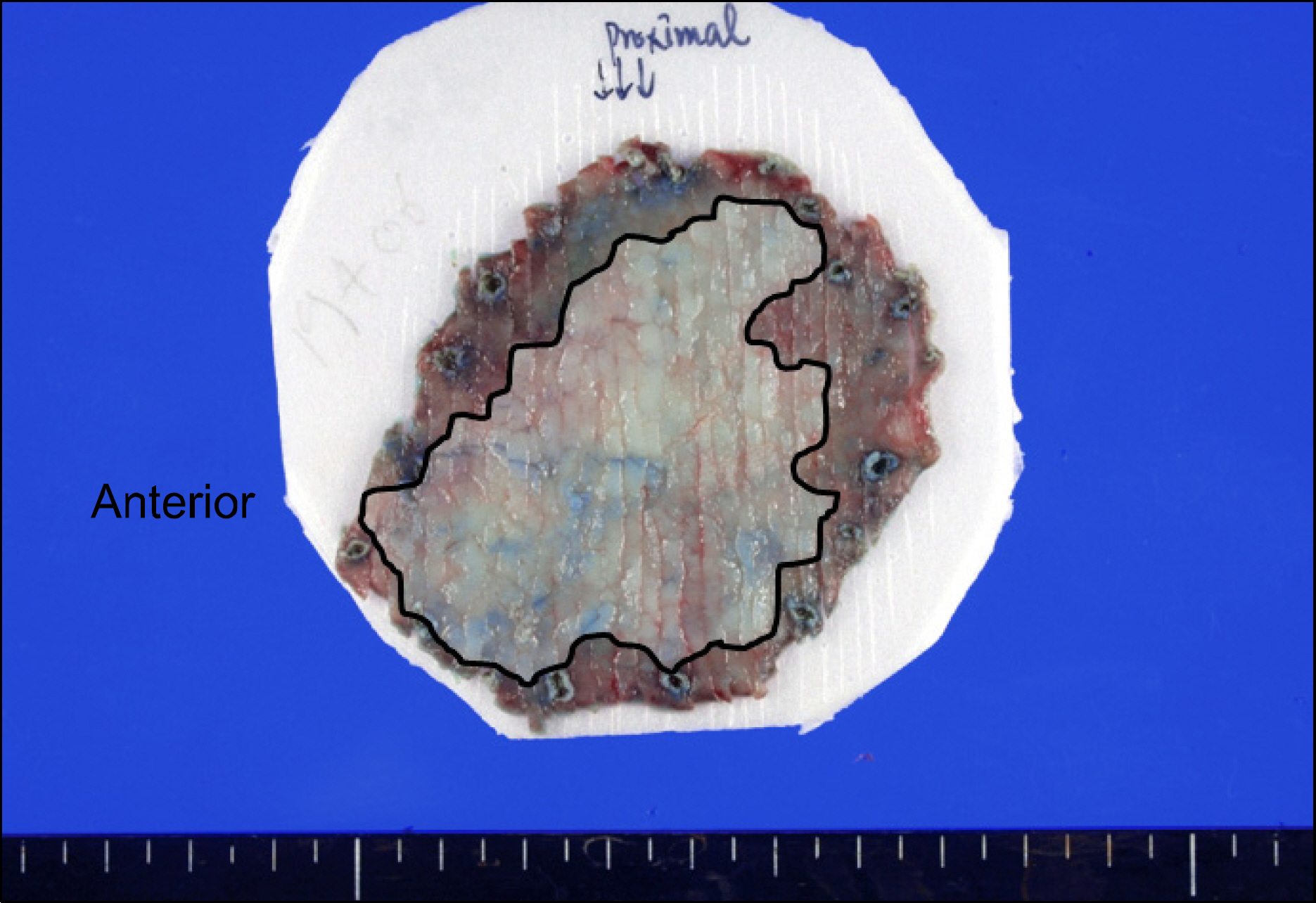
Fig. 6. Mapping of ESD specimen showed the involvement of anterior resection margin by type IIb early gastric carcinoma.
Cited by 1 articles
-
Clinical Outcomes of Endoscopic Submucosal Dissection for Undifferentiated or Submucosal Invasive Early Gastric Cancer
Pyung Gohn Goh, Hyun Yong Jeong, Min Jung Kim, Hyuk Soo Eun, Hye Jin Kim, Eui Sik Kim, Yun Jeung Kim, Soo Youn Lee, Hee Seok Moon, Eaum Seok Lee, Seok Hyun Kim, Jae Kyu Sung, Byung Seok Lee
Clin Endosc. 2011;44(2):116-122. doi: 10.5946/ce.2011.44.2.116.
Reference
-
1. Japanese Gastric Cancer Association. Treatment guideline of gastric cancer in Japan (Japanese). 2nd ed.Tokyo: Kanehara;2004.2. Japanese Gastric Cancer Association. Japanese classification of gastric carcinoma. 13th ed.Tokyo: Kanehara;1999.3. Kim WH, Park CH, Kim YB, et al. A standardized pathology report for gastric cancer. Korean J Pathol. 2005; 39:106–113.4. Lee SY, Park YS, Jang SJ, Oh ST, Huh JR. Differentiated intramucosal gastric carcinoma without ulceration showing extensive lymph node metastasis. Basic and Applied Pathology. 2010; 3:27–30.
Article5. Roh JH, Srivastava A, Lauwers GY, et al. Micropapillary carcinoma of stomach: a clinicopathologic and immunohistochemical study of 11 cases. Am J Surg Pathol. 2010; 34:1139–1146.6. Nakamura Y, Yasuoka H, Tsujimoto M, et al. Importance of lymph vessels in gastric cancer: a prognostic indicator in general and a predictor for lymph node metastasis in early stage cancer. J Clin Pathol. 2006; 59:77–82.
Article7. Kenkyūkai I. Japanese classification of gastric carcinoma. 1st ed.Tokyo: Kanehara & Co.;1995.8. Lauwers GY, Ban S, Mino M, et al. Endoscopic mucosal resection for gastric epithelial neoplasms: a study of 39 cases with emphasis on the evaluation of specimens and recommendations for optimal pathologic analysis. Mod Pathol. 2004; 17:2–8.
Article9. Ono H, Kondo H, Gotoda T, et al. Endoscopic mucosal resection for treatment of early gastric cancer. Gut. 2001; 48:225–229.
Article10. Park EH, Kang KT, Kim BH. The histologic discrepancy before and after endoscopic submucosal dissection of gastric adenoma and early gastric cancer. Korean J Gastrointest Endosc. 2007; 34:125–131.11. Kim YJ, Park JC, Kim JH, et al. Histologic diagnosis based on forceps biopsy is not adequate for determining endoscopic treatment of gastric adenomatous lesions. Endoscopy. 2010; 42:620–626.
Article12. Min BH, Kim KM, Kim ER, et al. Endoscopic and histo-pathological characteristics suggesting the presence of gastric mucosal high grade neoplasia foci in cases initially diagnosed as gastric mucosal low grade neoplasia by forceps biopsy in Korea. J Gastroenterol. 2010 Jul 31. [Epub ahead of print].
Article13. Schlemper RJ, Itabashi M, Kato Y, et al. Differences in diagnostic criteria for gastric carcinoma between Japanese and western pathologists. Lancet. 1997; 349:1725–1729.
Article14. Downs-Kelly E, Mendelin JE, Bennett AE, et al. Poor inter-observer agreement in the distinction of high-grade dysplasia and adenocarcinoma in pretreatment Barrett's esophagus biopsies. Am J Gastroenterol. 2008; 103:2333–2340.
Article15. Mita T, Shimoda T. Risk factors for lymph node metastasis of submucosal invasive differentiated type gastric carcinoma: clinical significance of histological heterogeneity. J Gastroenterol. 2001; 36:661–668.
Article16. Morita M, Kuwano H, Baba H, et al. Multifocal occurrence of gastric carcinoma in patients with a family history of gastric carcinoma. Cancer. 1998; 83:1307–1311.
Article17. Kosaka T, Miwa K, Yonemura Y, et al. A clinicopathologic study on multiple gastric cancers with special reference to distal gastrectomy. Cancer. 1990; 65:2602–2605.
Article
- Full Text Links
-
- Actions
-
Cited
- CITED
-
- Close
- Share
-
- Similar articles
-
- History and Development of Accessories for Endoscopic Submucosal Dissection
- How to Interpret the Pathological Report before and after Endoscopic Submucosal Dissection of Early Gastric Cancer
- Debates on Colorectal Endoscopic Submucosal Dissection - Traction for Effective Dissection: Gravity Is Enough
- Pathological Interpretation of Gastric Tumors in Endoscopic Submucosal Dissection
- A Case of Pneumorrhachis and Pneumoscrotum Following Colon Endoscopic Submucosal Dissection
