Vertebral osteophyte of pre-modern Korean skeletons from Joseon tombs
- Affiliations
-
- 1Department of Anatomy, Seoul National University College of Medicine, Seoul, Korea. drdoogi@snu.ac.kr
- 2Department of Internal Medicine, Seoul National University Boramae Medical Center, Seoul National University College of Medicine, Seoul, Korea.
- 3Department of Anatomy, Dankook University College of Medicine, Cheonan, Korea.
- 4Department of Anatomy, Ewha Womans University School of Medicine, Seoul, Korea.
- 5Institute of Forensic Medicine, Seoul National University, Seoul, Korea.
- KMID: 2133913
- DOI: http://doi.org/10.5115/acb.2012.45.4.274
Abstract
- Spinal osteophytic changes are known to be affected by differences in age, sex, population, and mechanical stress. We examined Joseon skeletons (n=87) to obtain vertebral osteophytosis data on a pre-modern Korean population. The mean osteophytic value (MOV) of vertebrae increased in the cervical-thoracic-lumbar order. More severe osteophytosis was found in the vertebrae (C5, T9, T10, and L4) farthest from the line of gravity, while the general pattern of vertebral osteophytosis appeared similar to those of previous reports on other skeletal series. More severe osteophytes were much more common in the males, possibly due to their engaging in more strenuous physical labor than that of females. We also observed MOV patterns seemingly unique to the Joseon people, and findings not typically reported in previous studies. Although a full explanation of the factors contributing to vertebral-osteophytic development in Joseon Koreans will require further studies, the present results are meaningful to anatomists and anthropologists interested in osteophytic patterns occurring in an East Asian population.
Keyword
MeSH Terms
Figure
-

Fig. 1 Examples of osteophyte grading in this study. Lumbar vertebrae in (A-E) represent grades 0-4 respectively. (A) Grade 0: no indication of osteophytosis. (B) Grade 1: an osteophyte with slight lipping (indicated by arrow) on the margin. (C) Grade 2: more lipping (arrow) visible on the margins. (D) Grade 3: advanced lipping with the free end curving in the direction of the closest intervertebral space (indicated by arrow). (E) Grade 4: osteophytes of two or more adjacent vertebrae fused together.

Fig. 2 (A) Mean osteophytic value in cervical, thoracic, and lumbar vertebrae of Joseon skeletons. (B) Mean osteophytic value at a glance from the cervical to lumbar spine.
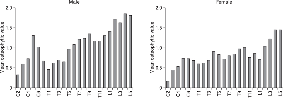
Fig. 3 Mean osteophytic value of Joseon skeletons at each spine by sex.
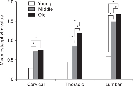
Fig. 4 Mean osteophytic value in the cervical, thoracic, and lumbar spines by age group. *P<0.05.
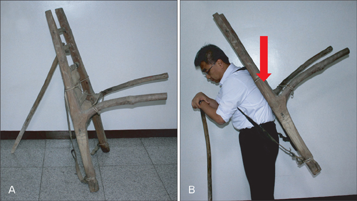
Fig. 5 (A) A-frame (Jige in Korean) traditionally used in premodern Korean society. (B) As a heavy burden could be carried using an A-frame, a heavy, strenuous load (indicated by arrow) occurred on the thoracic vertebrae.
Reference
-
1. van der Kraan PM, van den Berg WB. Osteophytes: relevance and biology. Osteoarthritis Cartilage. 2007. 15:237–244.2. Klaassen Z, Tubbs RS, Apaydin N, Hage R, Jordan R, Loukas M. Vertebral spinal osteophytes. Anat Sci Int. 2011. 86:1–9.3. Shore LR. Polyspondylitis marginalis osteophytica. Br J Surg. 1935. 22:850–863.4. Nathan H. Osteophytes of the vertebral column: an anatomical study of their development according to age, race, and sex with considerations as to their etiology and significance. J Bone Joint Surg Am. 1962. 44:243–268.5. Maat GJ, Mastwijk RW, Van der Velde EA. Skeletal distribution of degenerative changes in vertebral osteophytosis, vertebral osteoarthritis and DISH. Int J Osteoarchaeol. 1995. 5:289–298.6. Van der Merwe AE, Işcan MY, Lábbè EN. The pattern of vertebral osteophyte development in a South African population. Int J Osteoarchaeol. 2006. 16:459–464.7. Stewart TD. The rate of development of vertebral osteoarthritis in American whites and its significance in skeletal age identification. Leech. 1958. 28:144–151.8. Jurmain RD. The pattern of involvement of appendicular degenerative joint disease. Am J Phys Anthropol. 1980. 53:143–150.9. Ortner DJ, Putschar WG. Smithsonian Contributions to Anthropology No. 28. Identification of pathological conditions in human skeletal remains. 1981. Washington, DC: Smithsonian Institution Press.10. Krogman WM, İşan MY. The human skeleton in forensic medicine. 1986. Springfield: Charles C Thomas Publisher, Ltd..11. Jurmain R. Paleoepidemiology of a central California prehistoric population from Ca-Ala-329: II. Degenerative disease. Am J Phys Anthropol. 1990. 83:83–94.12. MacLaughlin SM, Oldale KN. Vertebral body diameters and sex prediction. Ann Hum Biol. 1992. 19:285–292.13. Bridges PS. Vertebral arthritis and physical activities in the prehistoric southeastern United States. Am J Phys Anthropol. 1994. 93:83–93.14. Lovell NC. Spinal arthritis and physical stress at Bronze Age Harappa. Am J Phys Anthropol. 1994. 93:149–164.15. Knüsel CJ, Göggel S, Lucy D. Comparative degenerative joint disease of the vertebral column in the medieval monastic cemetery of the Gilbertine priory of St. Andrew, Fishergate, York, England. Am J Phys Anthropol. 1997. 103:481–495.16. Taitz C. Osteophytosis of the cervical spine in South African blacks and whites. Clin Anat. 1999. 12:103–109.17. Sofaer Derevenski JR. Sex differences in activity-related osseous change in the spine and the gendered division of labor at Ensay and Wharram Percy, UK. Am J Phys Anthropol. 2000. 111:333–354.18. Rogers J, Waldron T, Dieppe P, Watt I. Arthropathies in palaeopathology: the basis of classification according to most probable cause. J Archaeol Sci. 1987. 14:179–193.19. O'Neill TW, McCloskey EV, Kanis JA, Bhalla AK, Reeve J, Reid DM, Todd C, Woolf AD, Silman AJ. The distribution, determinants, and clinical correlates of vertebral osteophytosis: a population based survey. J Rheumatol. 1999. 26(4):842–848.20. Weber J, Czarnetzki A, Spring A. Paleopathological features of the cervical spine in the early middle ages: natural history of degenerative diseases. Neurosurgery. 2003. 53:1418–1423.21. Stirland AJ, Waldron T. Evidence for activity related markers in the vertebrae of the crew of the Mary Rose. J Archaeol Sci. 1997. 24:329–335.22. Rojas-Sepúlveda C, Ardagna Y, Dutour O. Paleoepidemiology of vertebral degenerative disease in a Pre-Columbian Muisca series from Colombia. Am J Phys Anthropol. 2008. 135:416–430.23. Shimoda Y, Nagaoka T, Moromizato K, Sunagawa M, Hanihara T, Yoneda M, Hirata K, Ono H, Amano T, Fukumine T, Ishida H. Degenerative changes of the spine in people from prehistoric Okhotsk culture and two ancient human groups from Kanto and Okinawa, Japan. Anthropol Sci. 2012. 120:1–21.24. Lovejoy CO, Meindl RS, Pryzbeck TR, Mensforth RP. Chronological metamorphosis of the auricular surface of the ilium: a new method for the determination of adult skeletal age at death. Am J Phys Anthropol. 1985. 68:15–28.25. Rogers J, Waldron T. DISH and the monastic way of life. Int J Osteoarchaeol. 2001. 11:357–365.26. Steinbock RT. Paleopathological diagnosis and interpretations: bone diseases in ancient human populations. 1976. Springfield: Charles C. Thomas Publisher, Ltd..27. Snodgrass JJ. Sex differences and aging of the vertebral column. J Forensic Sci. 2004. 49:458–463.28. Kruskal WH, Wallis WA. Use of ranks in one-criterion variance analysis. J Am Stat Assoc. 1952. 47:583–621.29. Fisher RA. Statistical methods for research workers. 1954. Edinburgh: Oliver and Boyd.30. Levy LF. Porter's neck. Br Med J. 1968. 2:16–19.31. Hollinshead WH. Functional anatomy of the limbs and back. 1969. 3rd ed. Philadelphia: W. B. Saunders.32. Novak M, Šlaus M. Vertebral pathologies in two early modern period (16th-19th century) populations from Croatia. Am J Phys Anthropol. 2011. 145:270–281.33. Waldron T. The prevalence of, and the relationship between some spinal diseases in a human skeletal population from London. Int J Osteoarchaeol. 1991. 1:103–110.34. Shin MH, Yi YS, Bok GD, Lee EJ, Spigelman M, Park JB, Min SR, Shin DH. Peña PA, Martín CR, Rodríguez MA, editors. How did mummification occur in bodies buried in tombs with a lime soil mixture barrier during the Joseon Dynasty in Korea. Mummies and Science World Mummies Research. 2008. Santa Cruz de Tenerife: Academia Canaria de la Historia;105–113.35. Buckberry JL, Chamberlain AT. Age estimation from the auricular surface of the ilium: a revised method. Am J Phys Anthropol. 2002. 119:231–239.36. Murray KA, Murray T. A test of the auricular surface aging technique. J Forensic Sci. 1991. 36:1162–1169.37. Schmitt A. Age-at-death assessment using the os pubis and the auricular surface of the ilium: a test on an identified Asian sample. Int J Osteoarchaeol. 2004. 14:1–6.38. Chapman FH. Vertebral osteophytosis in prehistoric populations of central and southern Mexico. Am J Phys Anthropol. 1972. 36:31–38.39. Prescher A. Anatomy and pathology of the aging spine. Eur J Radiol. 1998. 27:181–195.40. Pearsall DJ, Reid JG. Line of gravity relative to upright vertebral posture. Clin Biomech. 1992. 7:80–86.
- Full Text Links
-
- Actions
-
Cited
- CITED
-
- Close
- Share
-
- Similar articles
-
- Stable isotope analysis of Joseon people skeletons from the cemeteries of Old Seoul City, the capital of Joseon Dynasty
- Construction of Medieval Skeleton Collections with Human Remains from Tombs of Goryeo Dynasty, Korea
- Harris lines observed in human skeletons of Joseon Dynasty, Korea
- Bone tumors in pre-modern skulls from human skeletal series of Joseon Dynasty
- Infection patterns of trematode parasites among Joseon people
