Bone tumors in pre-modern skulls from human skeletal series of Joseon Dynasty
- Affiliations
-
- 1Bioanthropology and Paleopathology Laboratory, Department of Anatomy, Seoul National University College of Medicine, Seoul, Korea.
- 2Department of Anatomy, Ewha Womans University School of Medicine, Seoul, Korea.
- 3Hangang Institute of Cultural Heritage, Seoul, Korea.
- 4Special Duty Professor for Archaeology, Industry Academic Cooperation Foundation of Sangmyung University, Cheonan, Korea.
- 5Department of Radiology, Seoul National University Hospital, Seongnam, Korea.
- 6Paleopathology Laboratory, Department of Anatomy, Dankook University College of Medicine, Cheonan, Korea. mjukim99@dankook.ac.kr
- KMID: 2424825
- DOI: http://doi.org/10.5115/acb.2015.48.3.213
Abstract
- To date, there are still very few reports on benign-tumor cases based on East Asian skeletal series, even though other regions and continents have been well represented. In our study on the Joseon Human Skeletal Series, we identified benign bone tumors in two skeletons (cases Nos. 75 and 96). Our radiological analyses showed both cases to be homogeneous sclerotic bone masses aligned with the cranial vault suture. In a subsequent series of differential diagnoses, we determined both cases to be osteoma, the most common bone-tumor type reported for archaeological samples. Our study is the osteoarchaeological basis for this, the first-ever report on benign bone neoplasm in a pre-modern East Asian population.
Keyword
MeSH Terms
Figure
-
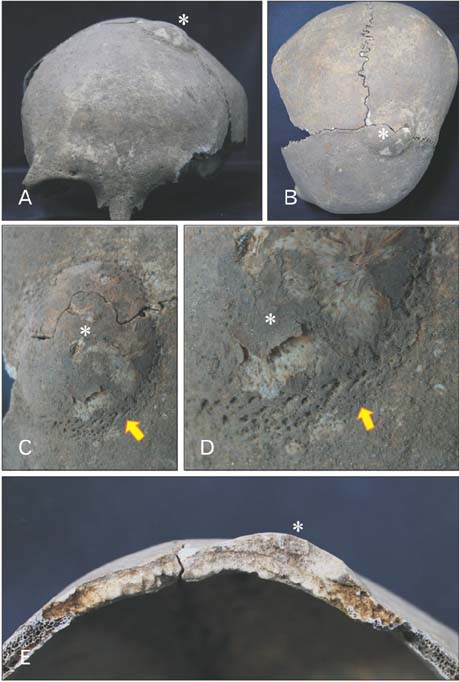
Fig. 1 Osteoma of case No. 75. (A) Frontal view of calvaria. (B) Superior view of calvaria. (A, B) Circular osteogenic mass (asterisks) located slightly aside from bregma, being in line with the left frontoparietal suture. (C) Well-circumscribed osteogenic mass is observed. (D) The magnified image of panel (C) showing porotic vascularized rim of a lesion (indicated by arrow). (E) Cross-sectional view of osteogenic mass showing external bulging of outer table of diploe with spongy bone. Note bone mass (asterisk) is identified in the outer table of skull.
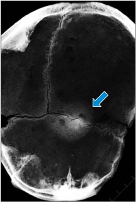
Fig. 2 Osteoma of case No. 75 on plain radiograph showing the extent of a lesion. Round homogenous mass with a smooth border (indicated by arrow) could be seen at the center of the image. Radiolucent areas were confirmed as arachnoid granulations.
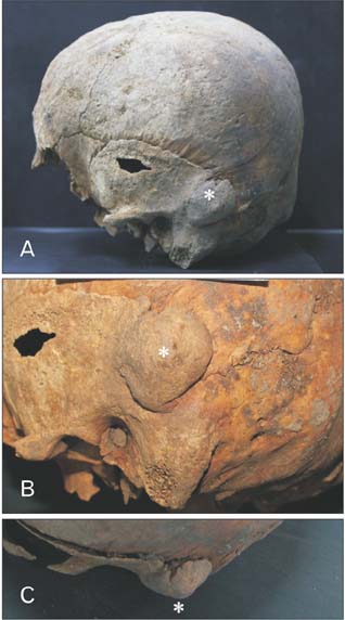
Fig. 3 Osteoma of case No. 96 in the left temporal bone. (A, B) Lateral view showing circular osteogenic mass (asterisks) lying in line with squamous suture. The hole shown in the squamous part of temporal bone is postmortem taphonomic change. Panel (C) is seen from above. Note that the margin of circular outgrowth mass above the external acoustic meatus is separated from the surrounding cortical bone.

Fig. 4 Osteoma of case No. 96 on plain radiograph showing well-circumscribed and homogeneous mass (indicated by arrows) with smooth border. (A) Lateral view. (B) Anterio-posterior view. (C) Magnified image of the mass.
Reference
-
1. Ortner DJ. Tumors and tumor-like lesions of bone. In : Ortner DJ, editor. Identification of Pathological Conditions in Human Skeletal Remains. 2nd ed. San Diego: Academic Press;2003. p. 503–544.2. Rogers L. Meningiomas in Pharaoh's people: hyperostosis in ancient Egyptian skulls. Br J Surg. 1949; 36:423.3. Gladykowska-Rzeczycka J, Urbanowicz M. Multiple exostoses in a skeleton from a prehistoric cemetary of the former population of Pruszcz Gdanski. Folia Morphol (Warsz). 1970; 29:317–329.4. Greenspan A. Benign bone-forming lesions: osteoma, osteoid osteoma, and osteoblastoma. Clinical, imaging, pathologic, and differential considerations. Skeletal Radiol. 1993; 22:485–500.5. Capasso LL. Antiquity of cancer. Int J Cancer. 2005; 113:2–13.6. Ruffer MA, Willmore JG. Studies in palaeopathology. Note on a tumour of the pelvis dating from Roman time (250 A.D.) and found in Egypt. J Pathol Bacteriol. 1913; 18:480–484.7. Oakley KP, Tobias PV. The Kanam jaw. Nature. 1960; 185:945–947.8. Sawyer DR, Wood NK, Allison MJ. An ancient "tumour" from pre-Columbian Chile. J Craniomaxillofac Surg. 1990; 18:136–138.9. Kim MJ, Oh CS, Lee IS, Lee BH, Choi JH, Lim DS, Yi YS, Han WJ, Kim YS, Bok GD, Lee SD, Shin DH. Human mummified brain from a medieval tomb with lime-soil mixture barrier of the Joseon Dynasty, Korea. Int J Osteoarchaeol. 2008; 18:614–623.10. Buikstra JE, Ubelaker DH. Standards for data collection from human skeletal remains. Arkansas Archaeological Survey Research Series, No. 44. Fayetteville, AR: Arkansas Archaeological Survey;1994.11. Lovejoy CO, Meindl RS, Pryzbeck TR, Mensforth RP. Chronological metamorphosis of the auricular surface of the ilium: a new method for the determination of adult skeletal age at death. Am J Phys Anthropol. 1985; 68:15–28.12. Schuller A. A short review of cranial hyperostoses. Acta radiol. 1950; 34:361–373.13. Dahlin DC, Johnson EW Jr. Giant osteoid osteoma. J Bone Joint Surg Am. 1954; 36:559–572.14. Jaffe HL. Benign osteoblastoma. Bull Hosp Joint Dis. 1956; 17:141–151.15. Vyhnánek L, Strouhal E, Nemmečková A. 'Kissing' osteochondroma: a case from Ancient Egypt. Int J Osteoarchaeol. 1999; 9:361–368.
- Full Text Links
-
- Actions
-
Cited
- CITED
-
- Close
- Share
-
- Similar articles
-
- The surface and the back of late Joseon medicine - Centered on medical knowledge system -
- Anthropometric Analysis of Buddha's Face of Korea and Joseon Dynasty in Buddhist Paintings and Comparison with the Beauty's Face of Joseon Dynasty
- Long bone fractures identified in the Joseon Dynasty human skeletons of Korea
- Processing Method & Distribution of Medicinal Plant Ginseng in Early Modern East Asia -Focusing on Ginseng as a Tribute Item of Joeseon to the Ming Dynasty-
- Anatomical Landmarks Used for Post-mortem Investigation Records Described in Jeungsu Muwonrok Eonhae of Joseon Dynasty Period
