Discal cysts of the cervical spine in two dogs
- Affiliations
-
- 1College of Veterinary Medicine, Kangwon National University, Chuncheon 24341, Korea.
- 2College of Veterinary Medicine, Chonbuk National University, Iksan 54596, Korea. kclee@chonbuk.ac.kr
- 3Office of Research Management, Korea University, Seoul 02841, Korea.
- KMID: 2133638
- DOI: http://doi.org/10.4142/jvs.2015.16.4.543
Abstract
- Discal cysts, which lie directly over intervertebral discs, are rare. Two old dogs with tetraparesis were referred to our facility. In both animals, magnetic resonance imaging revealed intraspinal extradural cystic mass lesions that were dorsal to degenerative intervertebral discs at the C3-C4 level. These lesions had low signal intensity on T1-weighted images, and high signal intensity on T2-weighted images. A ventral slot approach was used to perform surgical decompression, after which the symptoms improved remarkably. Discal cysts should be included in the differential diagnosis of dogs with cervical pain and tetraparesis. One effective treatment for discal cysts is surgical intervention.
Keyword
MeSH Terms
Figure
-
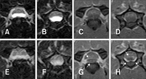
Fig. 1 Transverse T2-weighted (A and B), T1-weighted (C and D), FLAIR (E and F) and post-contrast T1-weighted (G and H) magnetic resonance images of Dog 1 at C3-C4. On T2-weighted (A and B) and FLAIR (E and F) images, a cyst is visible as a hyperintense structure on the ventral floor of the canal, compressing the spinal cord. On T1-weighted images (C and D), the cyst is slightly hypointense relative to the spinal cord. After administration of contrast medium (G and H), the cyst demonstrates capsular enhancement surrounding the hypointense fluid (arrows).

Fig. 2 Sagittal (A) and transverse (B) T2-weighted magnetic resonance images of Dog 2 at C3-C4. The discal cyst is hyperintense compared to the spinal cord and clearly visible in the ventral aspect of the canal (arrows). The spinal cord is deviated dorsally by the discal cyst on the sagittal and axial views (arrowhead).
Reference
-
1. Aydin S, Abuzayed B, Yildirim H, Bozkus H, Vural M. Discal cysts of the lumbar spine: report of five cases and review of the literature. Eur Spine J. 2010; 19:1621–1626.
Article2. Beltran E, Dennis R, Doyle V, de Stefani A, Holloway A, de Risio L. Clinical and magnetic resonance imaging features of canine compressive cervical myelopathy with suspected hydrated nucleus pulposus extrusion. J Small Anim Pract. 2012; 53:101–107.
Article3. Chiba K, Toyama Y, Matsumoto M, Maruiwa H, Watanabe M, Nishizawa T. Intraspinal cyst communicating with the intervertebral disc in the lumbar spine: discal cyst. Spine (Phila Pa 1976). 2001; 26:2112–2118.
Article4. Hamilton T, Glass E, Drobatz K, Agnello KA. Severity of spinal cord dysfunction and pain associated with hydrated nucleus pulposus extrusion in dogs. Vet Comp Orthop Traumatol. 2014; 27:313–318.
Article5. Kamishina H, Ogawa H, Katayama M, Yasuda J, Sato R, Tohyama K. Spontaneous regression of a cervical intraspinal cyst in a dog. J Vet Med Sci. 2010; 72:349–352.
Article6. Konar M, Lang J, Flüehmann G, Forterre F. Ventral intraspinal cysts associated with the intervertebral disc: magnetic resonance imaging observations in seven dogs. Vet Surg. 2008; 37:94–101.
Article7. Kono K, Nakamura H, Inoue Y, Okamura T, Shakudo M, Yamada R. Intraspinal extradural cysts communicating with adjacent herniated disks: imaging characteristics and possible pathogenesis. AJNR Am J Neuroradiol. 1999; 20:1373–1377.8. Lee HK, Lee DH, Choi CG, Kim SJ, Suh DC, Kahng SK, Roh SW, Rhim SC. Discal cyst of the lumbar spine: MR imaging features. Clin Imaging. 2006; 30:326–330.
Article9. Lowrie ML, Platt SR, Garosi LS. Extramedullary spinal cysts in dogs. Vet Surg. 2014; 43:650–662.
Article10. Penning VA, Benigni L, Steeves E, Cappello R. Imaging diagnosis-degenerative intraspinal cyst associated with an intervertebral disc. Vet Radiol Ultrasound. 2007; 48:424–427.
Article
