Displacement pattern of the anterior segment using antero-posterior lingual retractor combined with a palatal plate
- Affiliations
-
- 1Department of Orthodontics, School of Dentistry, Kyung Hee University, Seoul, Korea. acehyohyo@hanmail.net
- 2Division of Orthodontics, Department of Orofacial Science, University of California San Francisco, San Francisco, CA, USA.
- KMID: 2130591
- DOI: http://doi.org/10.4041/kjod.2015.45.6.289
Abstract
OBJECTIVE
To evaluate and compare the effects of two appliances on the en masse retraction of the anterior teeth anchored by temporary skeletal anchorage devices (TSADs).
METHODS
The sample comprised 46 nongrowing hyperdivergent adult patients who planned to undergo upper first premolar extraction using lingual retractors. They were divided into three groups, based on the lingual appliance used: the C-lingual retractor (CLR) group (group 1, n = 16) and two antero-posterior lingual retractor (APLR) groups (n = 30, groups 2 and 3). The APLR group was divided by the posterior tube angulation; posterior tube parallel to the occlusal plane (group 2, n = 15) and distally tipped tube (group 3, n = 15). A retrospective clinical investigation of the skeletal, dental, and soft tissue relationships was performed using lateral cephalometric radiographs obtained pretreatment and post en masse retraction of the anterior teeth.
RESULTS
All groups achieved significant incisor and canine retraction. The upper posterior teeth did not drift significantly during the retraction period. The APLR group had less angulation change in the anterior dentition, compared to the CLR group. By changing the tube angulation in the APLR, the intrusive force significantly increased in the distally tipped tube of group 3 patients and remarkably reduced the occlusal plane angle.
CONCLUSIONS
Compared to the CLR, the APLR provides better anterior torque control and canine tipping while achieving bodily translation. Furthermore, changing the tube angulation will affect the amount of incisor intrusion, even in patients with similar palatal vault depth, without the need for additional TSADs.
Figure
-

Figure 1 The occlusal diagrams and intraoral photos. A-C, The C-lingual retractor and D-F, the antero-posterior lingual retractor. A, No posterior orthodontic appliance is in place. B, Pretreatment and C, Post en masse retraction by the C-lingual retractor. D-F, The posterior teeth are splinted buccally and the guide bar and posterior tubes are in place. E, Pretreatment and F, Post en masse retraction by the antero-posterior lingual retractor.
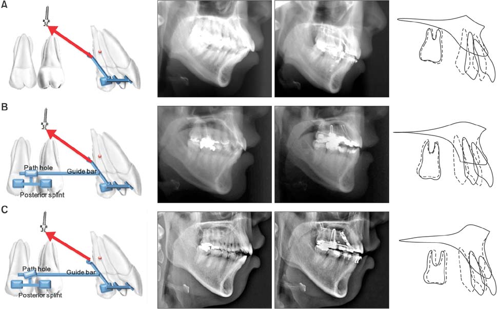
Figure 2 Schematic illustrations of the appliance in each group (lateral view; left column); lateral cephalograms at pretreatment and post en masse retraction (middle column); and maxillary superimposition (right column). A, The C-lingual retractor group (i.e., group 1) had clockwise rotation of the anterior segment during retraction. B, The antero-posterior lingual retractor with parallel tube group (i.e., group 2) had intrusion of the anterior segment with less clockwise moment. C, The antero-posterior lingual retractor with distally tipped tube group (i.e., group 3) had the greatest amount of intrusion on the anterior segment. Extrusion of the posterior segments did not occur in group 2 or group 3.
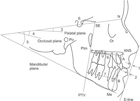
Figure 3 Soft tissue and skeletal cephalometric analysis. 1, The upper lip to the E line; 2, the lower lip to the E line; 3, the sella-nasion to the palatal plane angle (SN-PP); 4, the SN-anatomic occlusal plane angle (SN-Occ); 5, the SN to the mandibular plane angle (SN-Mn); 6, the distance between the pterygoid vertical plane and point A (PTV-A); 7, the distance between the pterygoid vertical plane and point B (PTV-B); and 8, the lower anterior face height (LAFH; ANS-Me). Refer Table 2 for definitions of the landmarks.
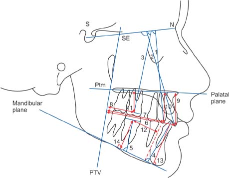
Figure 4 Dental cephalometric analysis, based on the angular and linear measurements. 1, The SN to the maxillary canine angle (SN-C); 2, the SN to the maxillary incisor angle (SN-U1); 3, the SN to the maxillary first molar angle (SN-U6); 4, the mandibular plane to mandibular incisor angle (MP-L1); 5, the mandibular plane to mandibular first molar angle (MP-L6); 6, the distance between the pterygoid vertical plane and the maxillary incisor tip (PTV-U1); 7, the distance between the pterygoid vertical plane and the maxillary canine tip (PTV-C); 8, the distance between the pterygoid vertical plane and the maxillary first molar centroid (PTV-U6); 9, the distance between the palatal plane and the maxillary incisor tip (PP-U1); 10, the distance between the palatal plane and the maxillary canine tip (PP-C); 11, the distance between the palatal plane and the maxillary first molar centroid (PP-U6); 12, the distance between the mandibular lingual cortex and the mandibular first molar centroid (LC-L6); 13, the distance between the mandibular plane and the mandibular incisor tip (MP-L1v); and 14, the distance between the mandibular plane and the mandibular first molar centroid (MP-L6v). Refer Table 2 for definitions of the landmarks.
Cited by 2 articles
-
Type of tooth movement during en masse retraction of the maxillary anterior teeth using labial versus lingual biocreative therapy in adults: A randomized clinical trial
Mais M. Sadek, Noha E. Sabet, Islam T. Hassan
Korean J Orthod. 2019;49(6):381-392. doi: 10.4041/kjod.2019.49.6.381.Effects of bracket slot size during
en-masse retraction of the six maxillary anterior teeth using an induction-heating typodont simulation system
Ji-Yong Kim, Won-Jae Yu, Prasad N. K. Koteswaracc, Hee-Moon Kyung
Korean J Orthod. 2017;47(3):158-166. doi: 10.4041/kjod.2017.47.3.158.
Reference
-
1. Hong RK, Heo JM, Ha YK. Lever-arm and mini-implant system for anterior torque control during retraction in lingual orthodontic treatment. Angle Orthod. 2005; 75:129–141.2. Lee EH, Yu HS, Lee KJ, Park YC. Three dimensional finite element analysis of continuous and segmented arches with use of orthodontic miniscrews. Korean J Orthod. 2011; 41:237–254.
Article3. Jeong GM, Sung SJ, Lee KJ, Chun YS, Mo SS. Finite-element investigation of the center of resistance of the maxillary dentition. Korean J Orthod. 2009; 39:83–94.
Article4. Chung KR, Oh MY, Ko SJ. Corticotomy-assisted orthodontics. J Clin Orthod. 2001; 35:331–339.5. Mo SS, Kim SH, Sung SJ, Chung KR, Chun YS, Kook YA, et al. Torque control during lingual anterior retraction without posterior appliances. Korean J Orthod. 2013; 43:3–14.
Article6. Chung KR, Kook YA, Kim SH, Mo SS, Jung JA. Class II malocclusion treated by combining a lingual retractor and a palatal plate. Am J Orthod Dentofacial Orthop. 2008; 133:112–123.
Article7. Nelson G, Ahn HW, Jeong SH, Kim JS, Kim SH, Chung KR. Three-dimensional retraction of anterior teeth with orthodontic miniplates in patients with temporomandibular disorder. Am J Orthod Dentofacial Orthop. 2012; 142:720–726.
Article8. Kim JS, Kim SH, Kook YA, Chung KR, Nelson G. Analysis of lingual en masse retraction combining a C-lingual retractor and a palatal plate. Angle Orthod. 2011; 81:662–669.
Article9. Kwon SY, Kim Y, Ahn HW, Kim KB, Chung KR, Kim Sunny SH. Computer-aided designing and manufacturing of lingual fixed orthodontic appliance using 2D/3D registration software and rapid prototyping. Int J Dent. 2014; 2014:164164.
Article10. Kwon SY, Ahn HW, Kim SH, Park YG, Chung KR, Paik CH, et al. Antero-posterior lingual sliding retraction system for orthodontic correction of hyperdivergent Class II protrusion. Head Face Med. 2014; 10:22.
Article11. Kim SH, Hwang YS, Ferreira A, Chung KR. Analysis of temporary skeletal anchorage devices used for enmasse retraction: a preliminary study. Am J Orthod Dentofacial Orthop. 2009; 136:268–276.
Article12. Jee JH, Ahn HW, Seo KW, Kim SH, Kook YA, Chung KR, et al. En-masse retraction with a preformed nickel-titanium and stainless steel archwire assembly and temporary skeletal anchorage devices without posterior bonding. Korean J Orthod. 2014; 44:236–245.
Article13. Melsen B. Biological reaction of alveolar bone to orthodontic tooth movement. Angle Orthod. 1999; 69:151–158.14. Ahn HW, Moon SC, Baek SH. Morphometric evaluation of changes in the alveolar bone and roots of the maxillary anterior teeth before and after en masse retraction using cone-beam computed tomography. Angle Orthod. 2013; 83:212–221.
Article15. Park JH, Tai K, Takagi M, Miyajima K, Kojima Y, Joo BH. Esthetic orthodontic treatment with a double J retractor and temporary anchorage devices. Am J Orthod Dentofacial Orthop. 2012; 141:796–805.
Article16. Hong RK, Lim SM, Heo JM, Baek SH. Orthodontic treatment of gummy smile by maxillary total intrusion with a midpalatal absolute anchorage system. Korean J Orthod. 2013; 43:147–158.
Article
- Full Text Links
-
- Actions
-
Cited
- CITED
-
- Close
- Share
-
- Similar articles
-
- Torque control during lingual anterior retraction without posterior appliances
- Three dimensional finite element analysis of continuous and segmented arches with use of orthodontic miniscrews
- A finite element analysis of the stress distribution and displacement in human maxilla to rapid palatal expansion
- Maxillary complete denture fabrication cases with posterior palatal seal considering palatal form and tissue displacement
- Pattern of microimplant displacement during maxillary skeletal expander treatment: A cone-beam computed tomography study
