Comparison of cone-beam computed tomography cephalometric measurements using a midsagittal projection and conventional two-dimensional cephalometric measurements
- Affiliations
-
- 1Department of Orthodontics, Gachon University Gil Medical Center, Incheon, Korea. orthodm@gilhospital.com
- KMID: 2130590
- DOI: http://doi.org/10.4041/kjod.2015.45.6.282
Abstract
OBJECTIVE
This study investigated whether it is possible to use a two-dimensional (2D) standard in three-dimensional (3D) analysis, by comparing the angles and lengths measured from a midsagittal projection in 3D cone-beam computed tomography (CBCT) with those measured by 2D lateral cephalometric radiography (LCR).
METHODS
Fifty patients who underwent both LCR and CBCT were selected as subjects. CBCT was reoriented in 3 different methods and the measuring-points were projected onto the midsagittal plane. Twelve angle values and 8 length values were measured on both LCR and CBCT and compared.
RESULTS
Repeated measures analysis of the variance revealed statistically significant differences in 7 angular and 5 linear measurements among LCR and 3 types of CBCT (p < 0.05). Of these 12 measurements, multiple comparisons showed that 6 measurements (ANB, AB to FH, IMPA, FMA, Co-Gn, Go-Me) were not significantly different in pairwise comparisons. LCR was significantly different from 3 types of CBCT in 3 angular (SN to FH, interincisal angle, FMIA) and 2 linear (S-Go, Co-ANS) measurements. The CBCT method was similar for all measurements, except for 1 linear measurement, i.e., S-N. However, the disparity between the mean values for all parameters was within the range of clinical measurement error.
CONCLUSIONS
3D-CBCT analysis, using midsagittal projection, is a useful method in which the 2D-LCR normative values can be used. Although the measurements changed with reorientation, these changes were not clinically significant.
Keyword
Figure
-
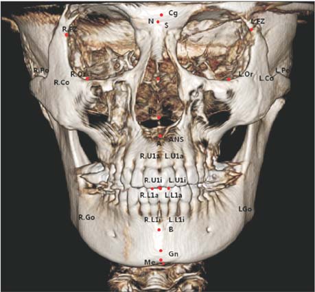
Figure 1 Landmarks used in three-dimensional cone-beam computed tomography analysis.
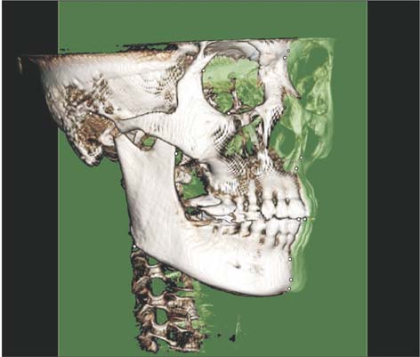
Figure 2 Midsagittal plane established by reorientation.
Cited by 5 articles
-
The genial tubercle: A prospective novel landmark for the diagnosis of mandibular asymmetry
Seung-Youp Lee, Dong-Soon Choi, Insan Jang, Geun-Su Song, Bong-Kuen Cha
Korean J Orthod. 2017;47(1):50-58. doi: 10.4041/kjod.2017.47.1.50.Skeletal and dentoalveolar changes after miniscrew-assisted rapid palatal expansion in young adults: A cone-beam computed tomography study
Jung Jin Park, Young-Chel Park, Kee-Joon Lee, Jung-Yul Cha, Ji Hyun Tahk, Yoon Jeong Choi
Korean J Orthod. 2017;47(2):77-86. doi: 10.4041/kjod.2017.47.2.77.Accuracy of three-dimensional cephalograms generated using a biplanar imaging system
Ha-Yeon Park, Jae-Seo Lee, Jin-Hyoung Cho, Hyeon-Shik Hwang, Kyung-Min Lee
Korean J Orthod. 2018;48(5):292-303. doi: 10.4041/kjod.2018.48.5.292.Comparison of three midsagittal planes for three-dimensional cone beam computed tomography head reorientation
Eon-Hwa Lee, Hyung-Seog Yu, Kee-Joon Lee, Sang-Sun Han, Hwi-Dong Jung, Chung-Ju Hwang
Korean J Orthod. 2020;50(1):3-12. doi: 10.4041/kjod.2020.50.1.3.Maxillomandibular arch width differences at estimated centers of resistance: Comparison between normal occlusion and skeletal Class III malocclusion
Yun-Jin Koo, Sung-Hwan Choi, Byeong-Tak Keum, Hyung-Seog Yu, Chung-Ju Hwang, Birte Melsen, Kee-Joon Lee
Korean J Orthod. 2017;47(3):167-175. doi: 10.4041/kjod.2017.47.3.167.
Reference
-
1. Broadbent BH. A new x-ray technique and its application to orthodontia. Angle Orthod. 1931; 1:45–66.2. Geelen W, Wenzel A, Gotfredsen E, Kruger M, Hansson LG. Reproducibility of cephalometric landmarks on conventional film, hardcopy, and monitor-displayed images obtained by the storage phosphor technique. Eur J Orthod. 1998; 20:331–340.
Article3. Yoon YJ, Kim KS, Hwang MS, Kim HJ, Choi EH, Kim KW. Effect of head rotation on lateral cephalometric radiographs. Angle Orthod. 2001; 71:396–403.4. Halazonetis DJ. From 2-dimensional cephalograms to 3-dimensional computed tomography scans. Am J Orthod Dentofacial Orthop. 2005; 127:627–637.
Article5. Park TJ, Lee SH, Lee KS. A method for mandibular dental arch superimposition using 3D cone beam CT and orthodontic 3D digital model. Korean J Orthod. 2012; 42:169–181.
Article6. Choi JH, Yu HS, Lee KJ, Park YC. Three-dimensional evaluation of maxillary anterior alveolar bone for optimal placement of miniscrew implants. Korean J Orthod. 2014; 44:54–61.
Article7. Pinsky HM, Dyda S, Pinsky RW, Misch KA, Sarment DP. Accuracy of three-dimensional measurements using cone-beam CT. Dentomaxillofac Radiol. 2006; 35:410–416.
Article8. Periago DR, Scarfe WC, Moshiri M, Scheetz JP, Silveira AM, Farman AG. Linear accuracy and reliability of cone beam CT derived 3-dimensional images constructed using an orthodontic volumetric rendering program. Angle Orthod. 2008; 78:387–395.
Article9. Brown AA, Scarfe WC, Scheetz JP, Silveira AM, Farman AG. Linear accuracy of cone beam CT derived 3D images. Angle Orthod. 2009; 79:150–157.
Article10. de Oliveira AE, Cevidanes LH, Phillips C, Motta A, Burke B, Tyndall D. Observer reliability of three-dimensional cephalometric landmark identification on cone-beam computerized tomography. Oral Surg Oral Med Oral Pathol Oral Radiol Endod. 2009; 107:256–265.
Article11. Kumar V, Ludlow JB, Mol A, Cevidanes L. Comparison of conventional and cone beam CT synthesized cephalograms. Dentomaxillofac Radiol. 2007; 36:263–269.
Article12. van Vlijmen OJ, Bergé SJ, Swennen GR, Bronkhorst EM, Katsaros C, Kuijpers-Jagtman AM. Comparison of cephalometric radiographs obtained from conebeam computed tomography scans and conventional radiographs. J Oral Maxillofac Surg. 2009; 67:92–97.
Article13. Grauer D, Cevidanes LS, Styner MA, Heulfe I, Harmon ET, Zhu H, et al. Accuracy and landmark error calculation using cone-beam computed tomography-generated cephalograms. Angle Orthod. 2010; 80:286–294.
Article14. Kumar V, Ludlow J, Soares Cevidanes LH, Mol A. In vivo comparison of conventional and cone beam CT synthesized cephalograms. Angle Orthod. 2008; 78:873–879.
Article15. Ramírez-Sotelo LR, Almeida S, Ambrosano GM, Bóscolo F. Validity and reproducibility of cephalometric measurements performed in full and hemifacial reconstructions derived from cone beam computed tomography. Angle Orthod. 2012; 82:827–832.
Article16. Zamora N, Llamas JM, Cibrián R, Gandia JL, Paredes V. Cephalometric measurements from 3D reconstructed images compared with conventional 2D images. Angle Orthod. 2011; 81:856–864.
Article17. Nalçaci R, Oztürk F, Sökücü O. A comparison of two-dimensional radiography and three-dimensional computed tomography in angular cephalometric measurements. Dentomaxillofac Radiol. 2010; 39:100–106.
Article18. Yitschaky O, Redlich M, Abed Y, Faerman M, Casap N, Hiller N. Comparison of common hard tissue cephalometric measurements between computed tomography 3D reconstruction and conventional 2D cephalometric images. Angle Orthod. 2011; 81:11–16.
Article19. Gribel BF, Gribel MN, Manzi FR, Brooks SL, McNamara JA Jr. From 2D to 3D: an algorithm to derive normal values for 3-dimensional computerized assessment. Angle Orthod. 2011; 81:3–10.
Article20. Lee JK, Jung PK, Moon CH. Three-dimensional cone beam computed tomographic image reorientation using soft tissues as reference for facial asymmetry diagnosis. Angle Orthod. 2014; 84:38–47.
Article21. Albarakati SF, Kula KS, Ghoneima AA. The reliability and reproducibility of cephalometric measurements: a comparison of conventional and digital methods. Dentomaxillofac Radiol. 2012; 41:11–17.
Article22. Damstra J, Fourie Z, Ren Y. Comparison between two-dimensional and midsagittal three-dimensional cephalometric measurements of dry human skulls. Br J Oral Maxillofac Surg. 2011; 49:392–395.
Article23. Olmez H, Gorgulu S, Akin E, Bengi AO, Tekdemir I, Ors F. Measurement accuracy of a computer-assisted three-dimensional analysis and a conventional twodimensional method. Angle Orthod. 2011; 81:375–382.
Article24. Chate RA. Cephalometric landmark identification within the petrous temporal region. Br J Orthod. 1987; 14:33–41.
Article25. Stabrun AE, Danielsen K. Precision in cephalometric landmark identification. Eur J Orthod. 1982; 4:185–196.26. Dibbets JM, Nolte K. Comparison of linear cephalometric dimensions in Americans of European descent (Ann Arbor, Cleveland, Philadelphia) and Americans of African descent (Nashville). Angle Orthod. 2002; 72:324–330.27. Cavalcanti MG, Haller JW, Vannier MW. Three-dimensional computed tomography landmark measurement in craniofacial surgical planning: experimental validation in vitro. J Oral Maxillofac Surg. 1999; 57:690–694.
Article
- Full Text Links
-
- Actions
-
Cited
- CITED
-
- Close
- Share
-
- Similar articles
-
- The reliability of the cephalogram generated from cone-beam CT
- Comparison of conventional lateral cephalograms with corresponding CBCT radiographs
- 3-Dimensional analysis for class III malocclusion patients with facial asymmetry
- The Reliability of 3D CT Analysis for Facial Asymmetry
- A study on the cephalometric changes by the displacement of the mandibular condyles
