Imaging Sci Dent.
2012 Dec;42(4):201-205. 10.5624/isd.2012.42.4.201.
Comparison of conventional lateral cephalograms with corresponding CBCT radiographs
- Affiliations
-
- 1Department of Oral and Maxillofacial Radiology, College of Dentistry, Yonsei University, Seoul, Korea. csp@yuhs.ac
- KMID: 1806784
- DOI: http://doi.org/10.5624/isd.2012.42.4.201
Abstract
- PURPOSE
This study was performed to assess the compatibility of cone beam computed tomography (CBCT) synthesized cephalograms with conventional cephalograms, and to find a method for obtaining normative values for three-dimensional (3D) assessments.
MATERIALS AND METHODS
The sample group consisted of 10 adults with normal occlusion and well-balanced faces. They were imaged using conventional and CBCT cephalograms. The CBCT cephalograms were synthesized from the CBCT data using OnDemand 3D software. Twenty-one angular and 12 linear measurements from each imaging modality were compared and analyzed using paired-t test.
RESULTS
The linear measurements between the two imaging modalities were not statistically different (p>0.05) except for the U1 to facial plane distance. The angular measurements between the two imaging modalities were not statistically different (p>0.05) with the exception of the gonial angle, ANB difference, and facial convexity.
CONCLUSION
Two-dimensional cephalometric norms could be readily used for 3D quantitative assessment, if corrected for lateral cephalogram distortion.
Figure
-

Fig. 1 Anatomic landmarks and planes used in the cephalometric analysis.
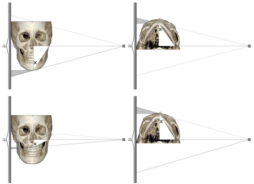
Fig. 2 Lateral cephalogram image construction diagram. 3D measurement=[(cephalometric measurement)-(cephalometric magnification)]/[cosine (X)].
Reference
-
1. Baumrind S, Frantz RC. The reliability of head film measurements. 1. Landmark identification. Am J Orthod. 1971. 60:111–127.2. Zamora N, Llamas JM, Cibrián R, Gandia JL, Paredes V. Cephalometric measurements from 3D reconstructed images compared with conventional 2D images. Angle Orthod. 2011. 81:856–864.
Article3. Oz U, Orhan K, Abe N. Comparison of linear and angular measurements using two-dimensional conventional methods and three-dimensional cone beam CT images reconstructed from a volumetric rendering program in vivo. Dentomaxillofac Radiol. 2011. 40:492–500.4. Kumar V, Ludlow J, Soares Cevidanes LH, Mol A. In vivo comparison of conventional and cone beam CT synthesized cephalograms. Angle Orthod. 2008. 78:873–879.
Article5. Broadbent BH. A new x-ray technique and its application to orthodontia. Angle Orthod. 1931. 1:45–66.6. Rousset MM, Simonek F, Dubus JP. A method for correction of radiographic errors in serial three-dimensional cephalometry. Dentomaxillofac Radiol. 2003. 32:50–59.
Article7. Farman AG, Scarfe WC. Development of imaging selection criteria and procedures should precede cephalometric assessment with cone-beam computed tomography. Am J Orthod Dentofacial Orthop. 2006. 130:257–265.
Article8. Cattaneo PM, Bloch CB, Calmar D, Hjortshøj M, Melsen B. Comparison between conventional and cone-beam computed tomography-generated cephalograms. Am J Orthod Dentofacial Orthop. 2008. 134:798–802.
Article9. Gribel BF, Gribel MN, Manzi FR, Brooks SL, McNamara JA Jr. From 2D to 3D: an algorithm to derive normal values for 3-dimensional computerized assessment. Angle Orthod. 2011. 81:3–10.
Article10. Kwon TG, Park HS, Ryoo HM, Lee SH. A comparison of craniofacial morphology in patients with and without facial asymmetry - a three-dimensional analysis with computed tomography. Int J Oral Maxillofac Surg. 2006. 35:43–48.11. Cevidanes LH, Bailey LJ, Tucker SF, Styner MA, Mol A, Phillips CL, et al. Three-dimensional cone-beam computed tomography for assessment of mandibular changes after orthognathic surgery. Am J Orthod Dentofacial Orthop. 2007. 131:44–50.
Article
- Full Text Links
-
- Actions
-
Cited
- CITED
-
- Close
- Share
-
- Similar articles
-
- Use of Reference Ear Plug to improve accuracy of lateral cephalograms generated from cone-beam computed tomography scans
- The comparative study of three-dimensional cephalograms to actual models and conventional lateral cephalograma in linear and angular measurements
- Accuracy of three-dimensional cephalograms generated using a biplanar imaging system
- Use of Head Posture Aligner to improve accuracy of frontal cephalograms generated from cone-beam CT scans
- Accuracy of virtual 3-dimensional cephalometric images constructed with 2-dimensional cephalograms using the biplanar radiography principle
