Comparison of Deep Anterior Lamellar Keratoplasty and Penetrating Keratoplasty for Keratoconus
- Affiliations
-
- 1Department of Ophthalmology, Samsung Medical Center, Sungkyunkwan University School of Medicine, Seoul, Korea. tychung@skku.edu
- KMID: 2127112
- DOI: http://doi.org/10.3341/jkos.2008.49.2.222
Abstract
-
PURPOSE: To compare the therapeutic outcomes after deep anterior lamellar keratoplasty (DALK) and penetrating keratoplasty (PKP) in patients with keratoconus.
METHODS
We retrospectively reviewed the clinical records of 57 patients diagnosed with keratoconus who had undergone DALK (19 eyes of 19 patients) and PKP (38 eyes of 38 patients) in Samsung medical center between January 1995 and January 2006.
RESULTS
The 19 and 38 patients with keratoconus who underwent DALK and PKP had mean ages of 25.3 (range: 17-46) and 26.2 (range: 12-51) years, respectively. These groups were followed up for mean times of 16.7 (range: 6-34) and 45.7 (range: 6-115) months after surgery, respectively. The DALK group showed significantly higher values of refractive power, central corneal thickness, and endothelial cell density, while two eyes (10.5%) in the DALK group developed stromal rejection, which resolved after steroid therapy. In the PKP group, eight eyes (21%) developed endothelial rejection, among whom one eye (2.6%) resulted in a graft failure of a patient who underwent re-PKP, and two eyes (5.3%) in the PKP group developed secondary glaucoma.
CONCLUSIONS
DALK should be considered as the primary surgical technique in keratoconus, because the visual outcome is comparable with PKP and it reduces severe complications such as secondary glaucoma and the risk of graft failure by preserving the corneal endothelium compared to PKP.
Keyword
MeSH Terms
Figure
-
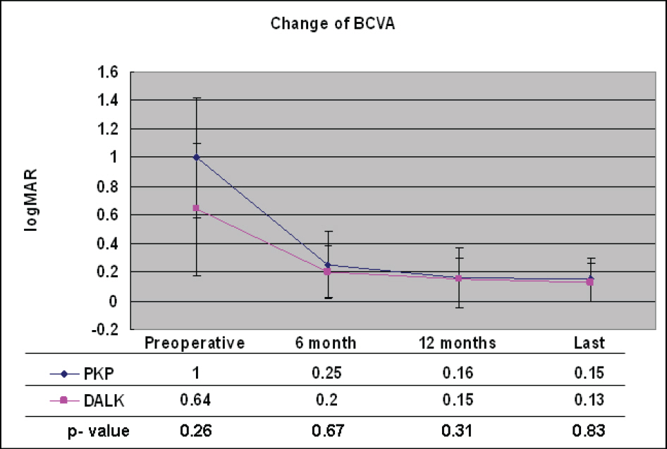
Figure 1. Change of logMAR best-corrected visual acuity (BCVA) after deep anterior lamellar keratoplasty (DALK) and penetrating keratoplasty (PKP). Mean BCVA preoperatively and up to the last follow-up after DALK (pink) and PKP (black) was provided. There were no statistical differences in BCVA between DALK and PKP groups throughout the observational period (Mann Whitney test).
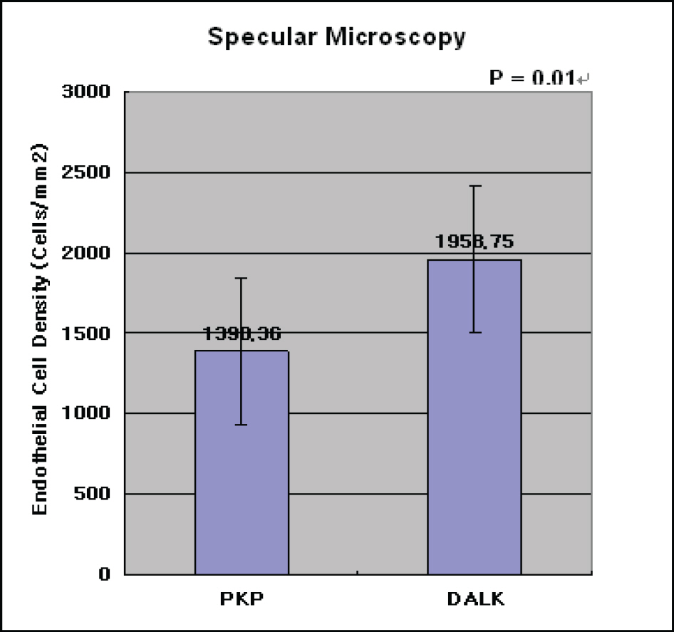
Figure 2. Difference of postoperative endothelial cell density between DALK (1958.6±455.1) and PKP (1390.4±460.2) group (p=0.01, Mann Whitney test). Specular microscopy was performed at median times of 26.0 months (DALK group) and 30.5 months (PKP group) postoperatively.
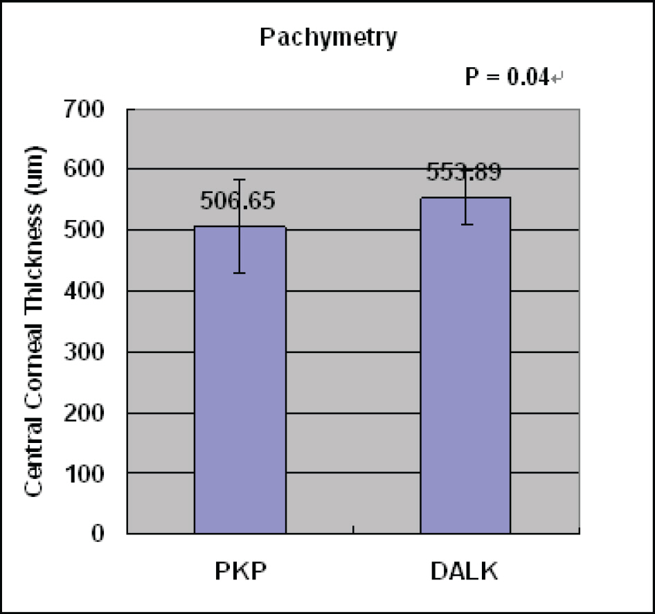
Figure 3. Difference of postoperative central corneal thickness between DALK (553.9±44,4) and PKP (506.7±77.4) group (p=0.04, Mann Whitney test). Pachymetry was performed at median times of 26.0 months (DALK group) and 30.5 months (PKP group) postoperatively.
Cited by 1 articles
-
Long-Term Effect and Safety of Contact Lenses for Keratoconus
Jong Heon Lee, Young Min Park, Young Kee Park, Jong Su Lee, Ji Eun Lee
J Korean Ophthalmol Soc. 2015;56(7):1006-1011. doi: 10.3341/jkos.2015.56.7.1006.
Reference
-
References
1. Edrington TB, Szczotka LB, Barr JT, et al. Rigid contact lens fitting relationships in keratoconus. Collaborative Longitudinal Evaluation of Keratoconus (CLEK) Study Group. Optom Vis Sci. 1999; 76:692–9.2. Wollensak G, Spoerl E, Seiler T. Riboflavin/ ultraviolet-a-induced collagen crosslinking for the treatment of keratoconus. Am J Ophthalmol. 2003; 135:620–7.3. Doh SH, Kim MS. Influence of Preoperative corneal thickness to postoperative astigmatism and endothelial cell in keratoconus penetrating keratoplasty. J Korean Ophthalmol Soc. 2005; 46:1978–82.4. Arentsen JJ. Corneal transplant allograft reaction: possible predisposing factors. Trans Am Ophthalmol Soc. 1983; 81:361–402.5. Ing JJ, Ing HH, Nelson LR, et al. Ten-year postoperative results of penetrating keratoplasty. Ophthalmology. 1998; 105:1855–65.
Article6. Tsubota K, Kaido M, Monden Y, et al. A new surgical technique for deep lamellar keratoplasty with single running suture adjustment. Am J Ophthalmol. 1998; 126:1–8.
Article7. Archila EA. Deep lamellar keratoplasty dissection of host tissue with intrastromal air injection. Cornea. 1984; 3:217–8.
Article8. Amayem AF, Anwar M. Fluid lamellar keratoplasty in keratoconus. Ophthalmology. 2000; 107:76–80.9. Sugita J, Kondo J. Deep lamellar keratoplasty with complete removal of pathological stroma for vision improvement. Br J Ophthalmol. 1997; 81:184–8.
Article10. Anwar M, Teichmann KD. Deep lamellar keratoplasty: surgical techniques for anterior lamellar keratoplasty with and without baring of Descemet's membrane. Cornea. 2002; 21:374–83.11. Fogla R, Padmanabhan P. Results of deep lamellar keratoplasty using the big-bubble technique in patients with keratoconus. Am J Ophthalmol. 2006; 141:254–9.
Article12. Luigi F, Gabriella P, Giorgio T. Clinical outcomes after deep anterior lamellar keratoplasty using the big-bubble technique in patients with keratoconus. Am J Ophthalmol. 2007; 143:117–24.13. Mo YH, Hahn TW, Kim JH. Comparison of lamellar and penetrating keratoplasty in keratoconus. J Korean Ophthalmol Soc. 1994; 35:908–14.14. Watson SL, Ramsay A, Dart JK, et al. Comparison of deep lamellarkeratoplasty and penetrating keratoplasty in patients with keratoconus. Ophthalmology. 2004; 111:1676–82.15. Krachmer JH, Mark JM, Edward JH. Cornea. 2nd ed.Vol. 1. Philadelphia: Elsevier;2005. p. 965–6.16. Maumenee AE. Clinical patterns of corneal graft failures. Ciba Foundation Symposium 15. Amsterdam/New York: Elsevier Publishing Company/Associated Scientific Publishers;1973. p. 5–23.17. Guo A, Ohia E, Xu JT, et al. Effects of anti-inflammatory and immunosuppressive drugs on the heterolamellar corneal transplantation in rabbits. Curr Eye Res. 1990; 9:749–57.
Article18. Soong HK, Katz DG, Farjo AA, et al. Central lamellar keratoplasty for optical indications. Cornea. 1999; 18:249–56.
Article19. Saini JS, Jain AK, Sukhija J, Saroha V. Indications and outcome of optical partial thickness lamellar keratoplasty. Cornea. 2003; 22:111–3.
Article20. Watson SL, Tuft SJ, Dart JK. Patterns of rejection after deep lamellar keratoplasty. Ophthalmology. 2006; 113:556–60.
Article21. Krachmer JH, Mark JM, Edward JH. Cornea. 2nd ed.Vol. 1. Philadelphia: Elsevier;2005. p. 1541–2.22. Bourne WM. Cellular changes in transplanted human corneas. Cornea. 2001; 20:560–9.
Article23. van Dooren BT, Mulder PG, Nieuwendaal CP, et al. Endothelial cell density after deep anterior lamellar keratoplasty (Melles technique). Am J Ophthalmol. 2004; 137:397–400.
Article
- Full Text Links
-
- Actions
-
Cited
- CITED
-
- Close
- Share
-
- Similar articles
-
- Clinical Evaluation of Full-thickness Deep Lamellar Keratoplasty
- Three Cases of Urrets-Zavalia Syndrome Following Deep Lamellar Keratoplasty (DLKP)
- A Case of Anterior Synechiolysis with Lamellar Corneal Dissection in Penetrating Keratoplasty
- Cataract Extraction after Penetrating Keratoplasty
- A Case of Double Descemet's Membrane after Penetrating Keratoplasty Converted from Deep Anterior Lamellar Keratoplasty
