Anthropometric analysis of maxillary anterior buccal bone of Korean adults using cone-beam CT
- Affiliations
-
- 1Department of Prosthodontics, College of Dentistry, Chosun University, Gwangju, Korea. jhajung@chosun.ac.kr
- KMID: 2118113
- DOI: http://doi.org/10.4047/jap.2010.2.3.92
Abstract
- PURPOSE
The aim of this study was to evaluate the thickness of buccal and palatal alveolar bone and buccal bony curvature below root apex in maxillary anterior teeth of Korean adults using Cone-beam CT images. MATERIAL AND METHODS: The 3D image was reconstructed with dicom file obtained through CBCT from 20 - 39 year old Korean subjects (n = 20). The thickness of buccal and palatal plate, root diameter, the buccal bony curvature angle below root apex and the distance from root apex to the deepest point of buccal bony curvature were measured on maxillary anterior teeth area using OnDemand3D program.
RESULTS
Mean thickness of buccal plate 3 mm below CEJ was 0.68 +/- 0.29 mm at central incisor, 0.76 +/- 0.59 mm at lateral incisor, and 1.07 +/- 0.80 mm at canine. Mean thickness of palatal plate 3 mm below CEJ was 1.53 +/- 0.55 mm of central incisor, 1.18 +/- 0.66 mm of lateral incisor, 1.42 +/- 0.77 mm of canine. Bucco-lingual diameter 3 mm below CEJ was 5.13 +/- 0.37 mm of central incisor, 4.58 +/- 0.46 mm of lateral incisor, and 5.93 +/- 0.47 mm of canine. Buccal bony curvature angle below root apex was 134.7 +/- 17.5degrees at central incisor, 151.0 +/- 13.9degrees at lateral incisor, 153.0 +/- 9.5degrees at canine. Distance between root apex and the deepest point of buccal bony curvature of central incisor was 3.67 +/- 1.28 mm at central incisor, 3.90 +/- 1.51 mm at lateral incisor, and 5.13 +/- 1.70 mm at canine.
CONCLUSION
Within the limitation of this study in Korean adults, the thickness of maxillary anterior buccal plate was very thin within 1mm and the thickness of palatal plate was thick, relatively. The buccal bony curvature below root apex of maxillary central incisor was higher than that of lateral incisor and canine and it seems that the buccal bony plate below root apex of central incisor is most curved.
Keyword
Figure
-
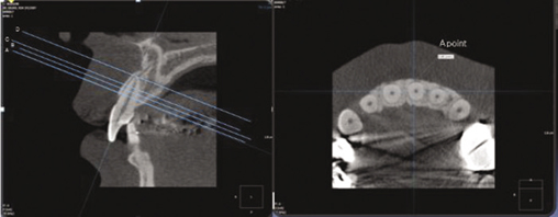
Fig. 1 Reference lines and measurement of the thickness of the buccal and palatal alveolar plates of each maxillary anterior tooth.
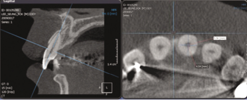
Fig. 2 Diameter of maxillary anterior teeth on reference line A.
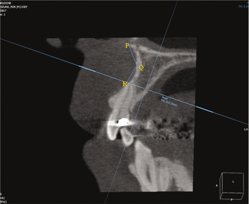
Fig. 3 Buccal bony curvature angle (∠PQR) below root apex of maxillary anterior teeth.

Fig. 4 Distance between root apex and the deepest point (Q) of buccal bony curvature.
Cited by 6 articles
-
Assessment of buccal bone thickness of aesthetic maxillary region: a cone-beam computed tomography study
Ramón Fuentes, Tania Flores, Pablo Navarro, Carlos Salamanca, Víctor Beltrán, Eduardo Borie
J Periodontal Implant Sci. 2015;45(5):162-168. doi: 10.5051/jpis.2015.45.5.162.New method of assessing the relationship between buccal bone thickness and gingival thickness
Yun-Jeong Kim, Ji-Man Park, Sungtae Kim, Ki-Tae Koo, Yang-Jo Seol, Yong-Moo Lee, In-Chul Rhyu, Young Ku
J Periodontal Implant Sci. 2016;46(6):372-381. doi: 10.5051/jpis.2016.46.6.372.Cone-beam computed tomographic analysis of the alveolar ridge profile and virtual implant placement for the anterior maxilla
Hyun-Chang Lim, Do-Uk Kang, Hyehyeon Baek, Ji-Youn Hong, Seung-Yun Shin, Jong-Hyuk Chung, Yeek Herr, Seung-Il Shin
J Periodontal Implant Sci. 2019;49(5):299-309. doi: 10.5051/jpis.2019.49.5.299.A novel classification of anterior alveolar arch forms and alveolar bone thickness: A cone-beam computed tomography study
Atcharee Bulyalert, Atiphan Pimkhaokham
Imaging Sci Dent. 2018;48(3):191-199. doi: 10.5624/isd.2018.48.3.191.Alveolar bone thickness and fenestration of incisors in untreated Korean patients with skeletal class III malocclusion: A retrospective 3-dimensional cone-beam computed tomography study
Song Hee Oh, Kyung-Yen Nahm, Seong-Hun Kim, Gerald Nelson
Imaging Sci Dent. 2020;50(1):9-14. doi: 10.5624/isd.2020.50.1.9.Accuracy of a direct drill-guiding system with minimal tolerance of surgical instruments used for implant surgery: a prospective clinical study
Du-Hyeong Lee, Seo-Young An, Min-Ho Hong, Kyoung-Bae Jeon, Kyu-Bok Lee
J Adv Prosthodont. 2016;8(3):207-213. doi: 10.4047/jap.2016.8.3.207.
Reference
-
1. Buser D, Martin W, Belser UC. Optimizing esthetics for implant restorations in the anterior maxilla: anatomic and surgical considerations. Int J Oral Maxillofac Implants. 2004. 19:43–61.2. Nevins M, Camelo M, De Paoli S, Friedland B, Schenk RK, Parma-Benfenati S, Simion M, Tinti C, Wagenberg B. A study of the fate of the buccal wall of extraction sockets of teeth with prominent roots. Int J Periodontics Restorative Dent. 2006. 26:19–29.3. Cardaropoli G, Araújo M, Lindhe J. Dynamics of bone tissue formation in tooth extraction sites. An experimental study in dogs. J Clin Periodontol. 2003. 30:809–818.4. Johnson K. A study of the dimensional changes occurring in the maxilla after tooth extraction. Part 2 : Closed face immediate denture treatment. Aust Dent J. 1964. 9:6–13.5. Araújo MG, Lindhe J. Dimensional ridge alterations following tooth extraction. An experimental study in the dog. J Clin Periodontol. 2005. 32:212–218.6. Schropp L, Wenzel A, Kostopoulos L, Karring T. Bone healing and soft tissue contour changes following single-tooth extraction: a clinical and radiographic 12-month prospective study. Int J Periodontics Restorative Dent. 2003. 23:313–323.7. Swasty D, Lee JS, Huang JC, Maki K, Gansky SA, Hatcher D, Miller AJ. Anthropometric analysis of the human mandibular cortical bone as assessed by cone-beam computed tomography. J Oral Maxillofac Surg. 2009. 67:491–500.8. Belser UC, Buser D, Hess D, Schmid B, Bernard JP, Lang NP. Aesthetic implant restorations in partially edentulous patientsa critical appraisal. Periodontol 2000. 1998. 17:132–150.9. Buser D, von Arx T. Surgical procedures in partially edentulous patients with ITI implants. Clin Oral Implants Res. 2000. 11:83–100.10. Langer B, Sullivan DY. Osseointegration: its impact on the interrelationship of periodontics and restorative dentistry. Part 3. Periodontal prosthesis redefined. Int J Periodontics Restorative Dent. 1989. 9:240–261.11. Becker W, Sennerby L, Bedrossian E, Becker BE, Lucchini JP. Implant stability measurements for implants placed at the time of extraction: a cohort, prospective clinical trial. J Periodontol. 2005. 76:391–397.12. London RM. The esthetic effects of implant platform selection. Compend Contin Educ Dent. 2001. 22:675–682.13. Kan JYK, Rungcharassaeng K. Immediate placement and provisionalization of maxillary anterior single implants: a surgical and prosthodontic rational. Pract Periodontics Aesthet Dent. 2000. 12:817–824.14. Smukler H, Castellucci F, Capri D. The role of the implant housing in obtaining aesthetics: generation of peri-implant gingivae and papillae-Part 1. Pract Proced Aesthet Dent. 2003. 15:141–149.15. Esposito M, Ekestubbe A, Gröndahl K. Radiological evaluation of marginal bone loss at tooth surfaces facing single Brånemark implants. Clin Oral Implants Res. 1993. 4:151–157.16. Garber DA, Salama MA, Salama H. Immediate total tooth replacement. Compend Contin Educ Dent. 2001. 22:210–216. 21817. Saadoun AP, Le Gall MG. Periodontal implications in implant treatment planning for aesthetic results. Pract Periodontics Aesthet Dent. 1998. 10:655–664.18. Gelb DA. Immediate implant surgery: three-year retrospective evaluation of 50 consecutive cases. Int J Oral Maxillofac Implants. 1993. 8:388–399.
- Full Text Links
-
- Actions
-
Cited
- CITED
-
- Close
- Share
-
- Similar articles
-
- Analysis of the root position and angulation of maxillary premolars in alveolar bone using cone-beam computed tomography
- Analysis of the root position of the maxillary incisors in the alveolar bone using cone-beam computed tomography
- Assessment of the relationship between the maxillary molars and adjacent structures using cone beam computed tomography
- A study on sagittal root position of maxillary anterior teeth in Korean
- Detection of maxillary second molar with two palatal roots using cone beam computed tomography: a case report
