Analysis of the root position of the maxillary incisors in the alveolar bone using cone-beam computed tomography
- Affiliations
-
- 1Department of Oral and Maxillofacial Radiology, School of Dentistry, Pusan National University, Yangsan, Korea.
- 2Department of Oral and Maxillofacial Radiology, Yonsei University College of Dentistry, Seoul, Korea. yona0668@yuhs.ac
- KMID: 2390083
- DOI: http://doi.org/10.5624/isd.2017.47.3.181
Abstract
- PURPOSE
The purpose of this study was to measure the buccal bone thickness and angulation of the maxillary incisors and to analyze the correlation between these parameters and the root position in the alveolar bone using cone-beam computed tomography (CBCT).
MATERIALS AND METHODS
CBCT images of 398 maxillary central and lateral incisors from 199 patients were retrospectively reviewed. The root position in the alveolar bone was classified as buccal, middle, or palatal, and the buccal type was further classified into subtypes I, II, and III. In addition, the buccolingual inclination of the tooth and buccal bone thickness were evaluated.
RESULTS
A majority of the maxillary incisors were positioned more buccally within the alveolar bone, and only 2 lateral incisors (0.5%) were positioned more palatally. The angulation of buccal subtype III was the greatest and that of the middle type was the lowest. Most of the maxillary incisors exhibited a thin facial bone wall, and the lateral incisors had a significantly thinner buccal bone than the central incisors. The buccal bone of buccal subtypes II and III was significantly thinner than that of buccal subtype I.
CONCLUSION
A majority of the maxillary incisor roots were positioned close to the buccal cortical plate and had a thin buccal bone wall. Significant relationships were observed between the root position in the alveolar bone, the angulation of the tooth in the alveolar bone, and buccal bone thickness. CBCT analyses of the buccal bone and sagittal root position are recommended for the selection of the appropriate treatment approach.
Keyword
MeSH Terms
Figure
-

Fig. 1 The root position of the incisors in the alveolar bone is classified as the buccal, middle, or palatal type. A. Buccal type: the apical point of the incisor is within the buccal third of the alveolar bone and the root is closer to the buccal bone wall. B. Middle type: the apical point of the incisor is within the middle third of the alveolar bone and the buccal and palatal bone walls are approximately equal in thickness. C. Palatal type: the apical point of the incisor is within the palatal third of the alveolar bone and the root is closer to the palatal bone wall.
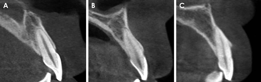
Fig. 2 The buccal type is further classified as follows. A. Subtype I: the incisor root is covered by the buccal bone wall, and the bone thickness increases toward the apex. B. Subtype II: the incisor root is covered by a thinner buccal bone wall than in subtype I and the bone thickness does not noticeably increase toward the apex that is covered by the bone tissue in the long axis of the tooth. C. Subtype III: the axis of the apex is angulated very buccally and the apex is not covered by the bone tissue in the long axis of the tooth.

Fig. 3 The angle between the long axis of the tooth and the long axis of the corresponding alveolar bone is measured.
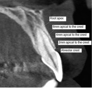
Fig. 4 The thickness of the buccal bone is measured at the alveolar crest; 2, 4, and 6 mm apical to the alveolar crest; and at the root apex.
Cited by 1 articles
-
Influence of the anterior arch shape and root position on root angulation in the maxillary esthetic area
Suweera Petaibunlue, Pravej Serichetaphongse, Atiphan Pimkhaokham
Imaging Sci Dent. 2019;49(2):123-130. doi: 10.5624/isd.2019.49.2.123.
Reference
-
1. Chan HL, Garaicoa-Pazmino C, Suarez F, Monje A, Benavides E, Oh TJ, et al. Incidence of implant buccal plate fenestration in the esthetic zone: a cone beam computed tomography study. Int J Oral Maxillofac Implants. 2014; 29:171–177.
Article2. Lau SL, Chow J, Li W, Chow LK. Classification of maxillary central incisors-implications for immediate implant in the esthetic zone. J Oral Maxillofac Surg. 2011; 69:142–153.
Article3. Wang HM, Shen JW, Yu MF, Chen XY, Jiang QH, He FM. Analysis of facial bone wall dimensions and sagittal root position in the maxillary esthetic zone: a retrospective study using cone beam computed tomography. Int J Oral Maxillofac Implants. 2014; 29:1123–1129.
Article4. Braut V, Bornstein MM, Belser U, Buser D. Thickness of the anterior maxillary facial bone wall-a retrospective radiographic study using cone beam computed tomography. Int J Periodontics Restorative Dent. 2011; 31:125–131.5. Kan JY, Roe P, Rungcharassaeng K, Patel RD, Waki T, Lozada JL, et al. Classification of sagittal root position in relation to the anterior maxillary osseous housing for immediate implant placement: a cone beam computed tomography study. Int J Oral Maxillofac Implants. 2011; 26:873–876.6. Araújo MG, Lindhe J. Dimensional ridge alterations following tooth extraction. An experimental study in the dog. J Clin Periodontol. 2005; 32:212–218.
Article7. Grunder U, Gracis S, Capelli M. Influence of the 3-D bone-to-implant relationship on esthetics. Int J Periodontics Restorative Dent. 2005; 25:113–119.8. Sung CE, Cochran DL, Cheng WC, Mau LP, Huang PH, Fan WH, et al. Preoperative assessment of labial bone perforation for virtual immediate implant surgery in the maxillary esthetic zone: a computer simulation study. J Am Dent Assoc. 2015; 146:808–819.9. El Nahass H, Naiem SN. Analysis of the dimensions of the labial bone wall in the anterior maxilla: a cone-beam computed tomography study. Clin Oral Implants Res. 2015; 26:e57–e61.
Article10. Rugani P, Kirnbauer B, Arnetzl GV, Jakse N. Cone beam computerized tomography: basics for digital planning in oral surgery and implantology. Int J Comput Dent. 2009; 12:131–145.11. Spector L. Computer-aided dental implant planning. Dent Clin North Am. 2008; 52:761–775.
Article12. Mandelaris GA, Rosenfeld AL. The expanding influence of computed tomography and the application of computer-guided implantology. Pract Proced Aesthet Dent. 2008; 20:297–305.13. Guerrero ME, Noriega J, Jacobs R. Preoperative implant planning considering alveolar bone grafting needs and complication prediction using panoramic versus CBCT images. Imaging Sci Dent. 2014; 44:213–220.
Article14. Xu D, Wang Z, Sun L, Lin Z, Wan L, Li Y, et al. Classification of the root position of the maxillary central incisors and its clinical significance in immediate implant placement. Implant Dent. 2016; 25:520–524.
Article15. Khoury J, Ghosn N, Mokbel N, Naaman N. Buccal bone thickness overlying maxillary anterior teeth: a clinical and radiographic prospective human study. Implant Dent. 2016; 25:525–531.16. Chung SH, Park YS, Chung SH, Shon WJ. Determination of implant position for immediate implant placement in maxillary central incisors using palatal soft tissue landmarks. Int J Oral Maxillofac Implants. 2014; 29:627–633.
Article17. Vera C, De Kok IJ, Reinhold D, Limpiphipatanakorn P, Yap AK, Tyndall D, et al. Evaluation of buccal alveolar bone dimension of maxillary anterior and premolar teeth: a cone beam computed tomography investigation. Int J Oral Maxillofac Implants. 2012; 27:1514–1519.18. Huynh-Ba G, Pjetursson BE, Sanz M, Cecchinato D, Ferrus J, Lindhe J, et al. Analysis of the socket bone wall dimensions in the upper maxilla in relation to immediate implant placement. Clin Oral Implants Res. 2010; 21:37–42.
Article19. Nowzari H, Molayem S, Chiu CH, Rich SK. Cone beam computed tomographic measurement of maxillary central incisors to determine prevalence of facial alveolar bone width ≥2 mm. Clin Implant Dent Relat Res. 2012; 14:595–602.20. Januário AL, Duarte WR, Barriviera M, Mesti JC, Araújo MG, Lindhe J. Dimension of the facial bone wall in the anterior maxilla: a cone-beam computed tomography study. Clin Oral Implants Res. 2011; 22:1168–1171.
Article21. Zekry A, Wang R, Chau AC, Lang NP. Facial alveolar bone wall width - a cone-beam computed tomography study in Asians. Clin Oral Implants Res. 2014; 25:194–206.
Article22. Miyamoto Y, Obama T. Dental cone beam computed tomography analyses of postoperative labial bone thickness in maxillary anterior implants: comparing immediate and delayed implant placement. Int J Periodontics Restorative Dent. 2011; 31:215–225.23. Buser D, Chen ST, Weber HP, Belser UC. Early implant placement following single-tooth extraction in the esthetic zone: biologic rationale and surgical procedures. Int J Periodontics Restorative Dent. 2008; 28:441–451.24. Ferrus J, Cecchinato D, Pjetursson EB, Lang NP, Sanz M, Lindhe J. Factors influencing ridge alterations following immediate implant placement into extraction sockets. Clin Oral Implants Res. 2010; 21:22–29.
Article
- Full Text Links
-
- Actions
-
Cited
- CITED
-
- Close
- Share
-
- Similar articles
-
- Analysis of the root position and angulation of maxillary premolars in alveolar bone using cone-beam computed tomography
- Alveolar bone thickness around maxillary central incisors of different inclination assessed with cone-beam computed tomography
- Assessment of the relationship between the maxillary molars and adjacent structures using cone beam computed tomography
- Associations among the anterior maxillary dental arch form, alveolar bone thickness, and the sagittal root position of the maxillary central incisors in relation to immediate implant placement: A cone-beam computed tomography analysis
- Retrospective Analysis of Incisor Root Resorption Associated with Impacted Maxillary Canines
