Post traumatic malocclusion and its prosthetic treatment
- Affiliations
-
- 1Department of Prosthodontics and Dental Research Institute, School of Dentistry, Seoul National University, Seoul, Korea. ksy0617@snu.ac.kr
- KMID: 2118112
- DOI: http://doi.org/10.4047/jap.2010.2.3.88
Abstract
- Mandible fractures belong to the most common fractures encountered in maxillofacial trauma. Because mandible is such a unique structure with hinge joint and masticatory muscles attached to the body of mandible, attention must be paid to avoid displacement during treatment. Displacement during fracture reduction leads to malocclusion. Many TMJs function with complete comfort and apparent normalcy in adapted centric posture, even though they have undergone deformation caused by trauma. This clinical report describes the patient with post traumatic malocclusion and its prosthetic treatment. His fractured mandible was openly reduced in changed position, as a result his occlusion has been changed. He was treated by prosthetic method in so-called adapted centric posture.
Keyword
MeSH Terms
Figure
-
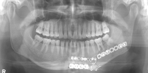
Fig. 1 Panoramic radiograph when the patient first visited Department of Orthodontics at the Seoul National University Dental Hospital in July 2007.

Fig. 2 Panoramic radiograph after open reduction surgery at the Department of Oral and Maxillofacial Surgery in September 2007.

Fig. 3 Teeth in maximum intercuspation when the patient first visited Department of Prosthodontics in July 2008. A: right lateral view, B: frontal view, C: left lateral view.

Fig. 4 Articulated study models in centric relation. A: right lateral view, B: frontal view, C: left lateral view.
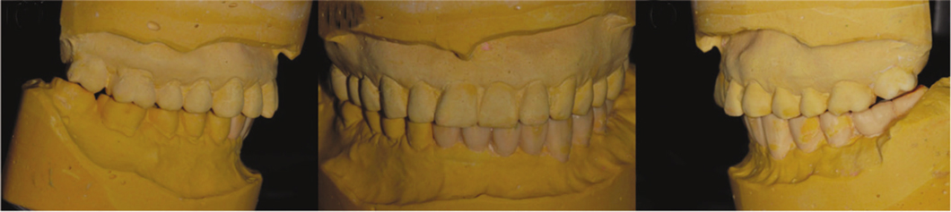
Fig. 5 Articulated diagnostic wax up models. A: right lateral view, B: frontal view, C: left lateral view.

Fig. 6 Teeth in maximum cuspationtion (= centric relation), post treatment. A: right lateral view, B: frontal view, C: left lateral view.
Reference
-
1. Motamedi MH. An assessment of maxillofacial fractures: a 5-year study of 237 patients. J Oral Maxillofac Surg. 2003. 61:61–64.2. Marciani RD, Carlson ER, Braun TW. Oral and maxiollofacial surgery II. 2009. 2nd ed. St. Louis: Saunders/Elsevier;139–143.3. Fordyce AM, Lalani Z, Songra AK, Hildreth AJ, Carton AT, Hawkesford JE. Intermaxillary fixation is not usually necessary to reduce mandibular fractures. Br J Oral Maxillofac Surg. 1999. 37:52–57.4. Renton TF, Wiesenfeld D. Mandibular fracture osteosynthesis: a comparison of three techniques. Br J Oral Maxillofac Surg. 1996. 34:166–173.5. Dawson PE. New definition for relating occlusion to varying conditions of the temporomandibular joint. J Prosthet Dent. 1995. 74:619–627.6. Dawson PE. Functional occlusion: From TMJ to smile design. 2007. 1st ed. St. Louis: MO: Mosby;69–74.7. Dawson PE. Functional occlusion from TMJ to smile design. 2007. St. Louis: Mosby Elsevier;345–348.
- Full Text Links
-
- Actions
-
Cited
- CITED
-
- Close
- Share
-
- Similar articles
-
- Occlusal rehabilitation of post-traumatic malocclusion patient after reduction of panfacial fracture, using selective occlusal adjustment and implant prostheses on centric relation: a case report
- The surgical correction of post-traumatic malocclusion
- A roentgenocephalometric study on soft tissue profile changes in pre-post treatment of Angle's Class II division I malocclusion
- Le Fort I osteotomy as treatment for traumatic class III malocclusion caused by Le Fort III fracture: A case report
- Post-Traumatic Stress Disorder, Depression and Anxiety among North Korean Refugees: A Meta-Analysis
