Occlusal rehabilitation of post-traumatic malocclusion patient after reduction of panfacial fracture, using selective occlusal adjustment and implant prostheses on centric relation: a case report
- Affiliations
-
- 1Department of Prosthodontics, School of Dentistry, Jeonbuk National University, Research Institute of Clinical Medicine of Jeonbuk National university - Biomedical Research Institute of Jeonbuk National University Hospital, Jeonju, Republic of Korea
- 2Department of Dentistry, Daejeon Konyang Medical Center, Konyang University, School of Medicine, Daejeon, Republic of Korea
- KMID: 2550429
- DOI: http://doi.org/10.14368/jdras.2023.39.4.204
Abstract
- Invasive or non-invasive reduction of fractures could be conducted as treatments of traumatic maxillofacial bone fractures. But when suboptimal reduction or malunion of maxillofacial bone fracture occurs, malocclusion could occur as a result of the lost relationship of the mandible and midface. This malocclusion is called post-traumatic malocclusion and orthognathic surgery, orthodontic treatment, selective grinding and prosthetic reconstruction are suggested as treatments for post-traumatic malocclusion after securement of stable TMJ. Stable TMJ is essential for occlusal rehabilitation to prevent occlusal change and relapse of malocclusion. Centric relation and adapted centric posture are suggested as start points of occlusal rehabilitation because they are most stable TMJ position. This case report presents a case in which post-traumatic malocclusion occurred after reduction of panfacial fracture. To rehabilitate full mouth occlusion, selective grinding and prosthetic reconstruction of implant supported fixed prostheses were conducted in centric relation and showed satisfying results in functional and occlusal aspects.
Keyword
Figure
-
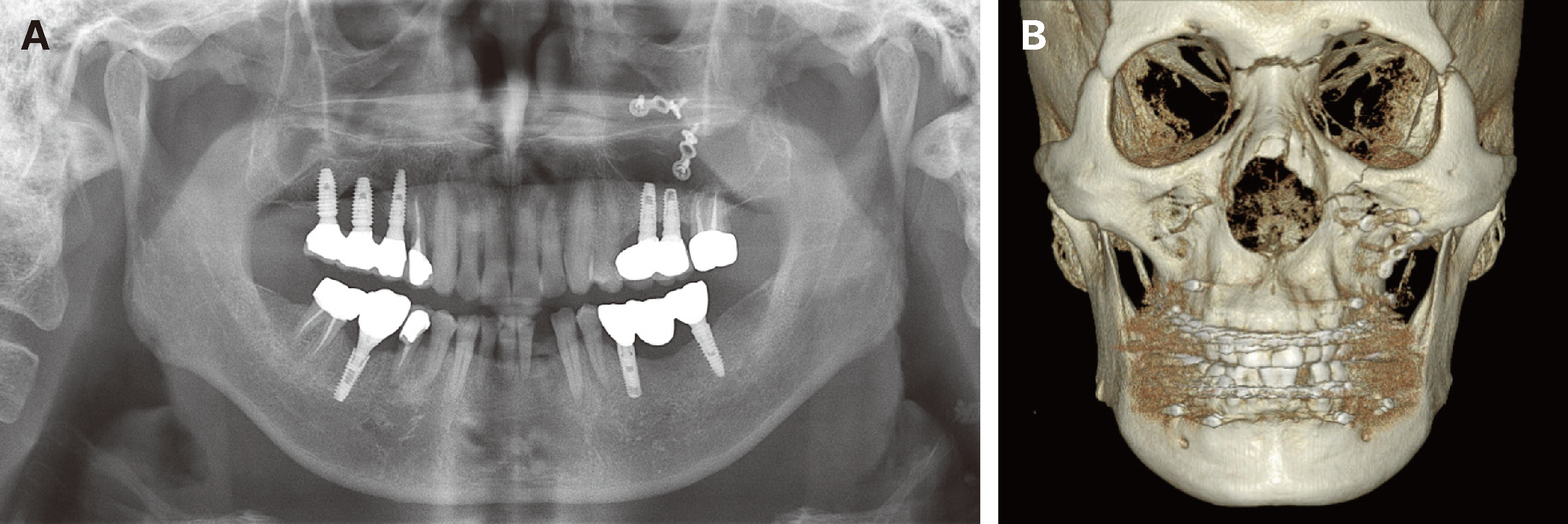
Fig. 1 Radiographic examinations after reduction of panfacial fractures. (A) Panoramic radiograph, (B) 3D image of cone beam CT.

Fig. 2 Intraoral view of patient. (A) Maxillary occlusal view, (B) Right lateral view, (C) Frontal view, (D) Left lateral view, (E) Mandibular occlusal view.
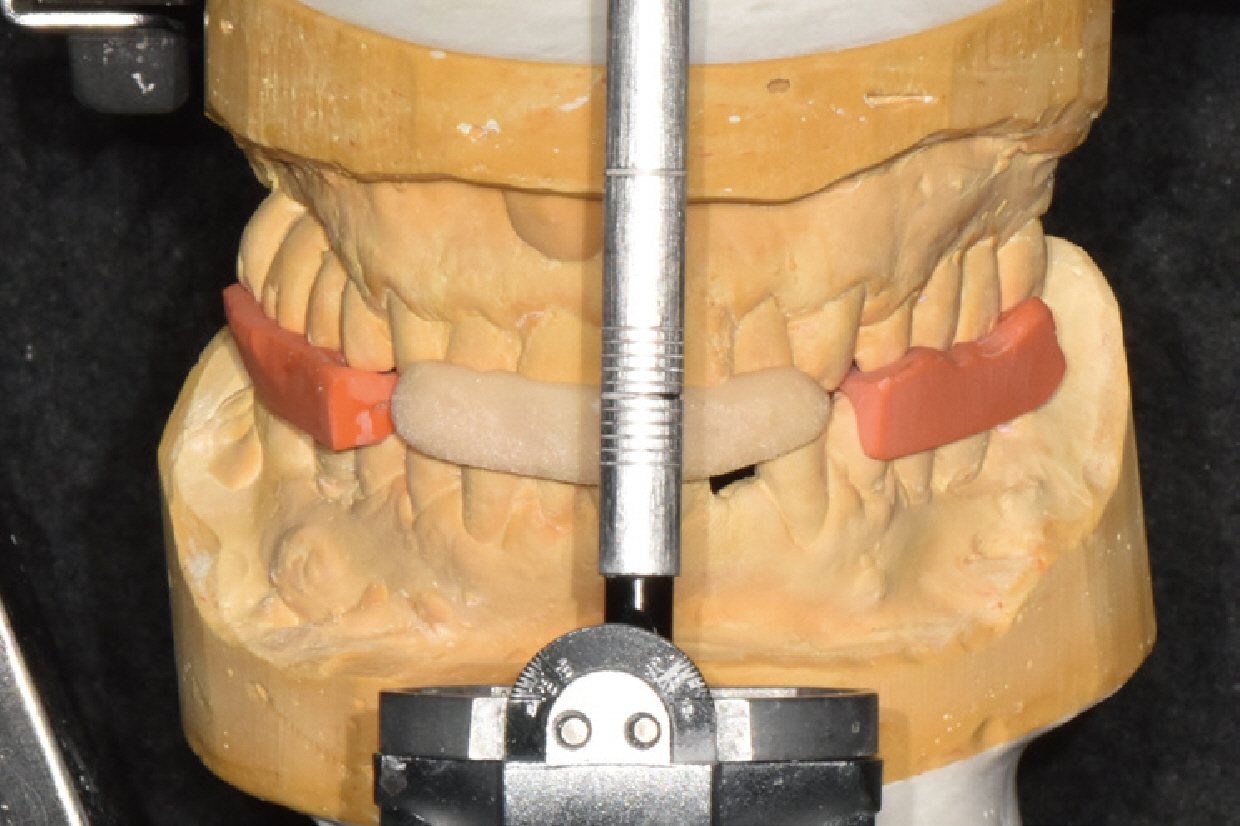
Fig. 3 Diagnostic mounting of diagnostic casts on centric relation.
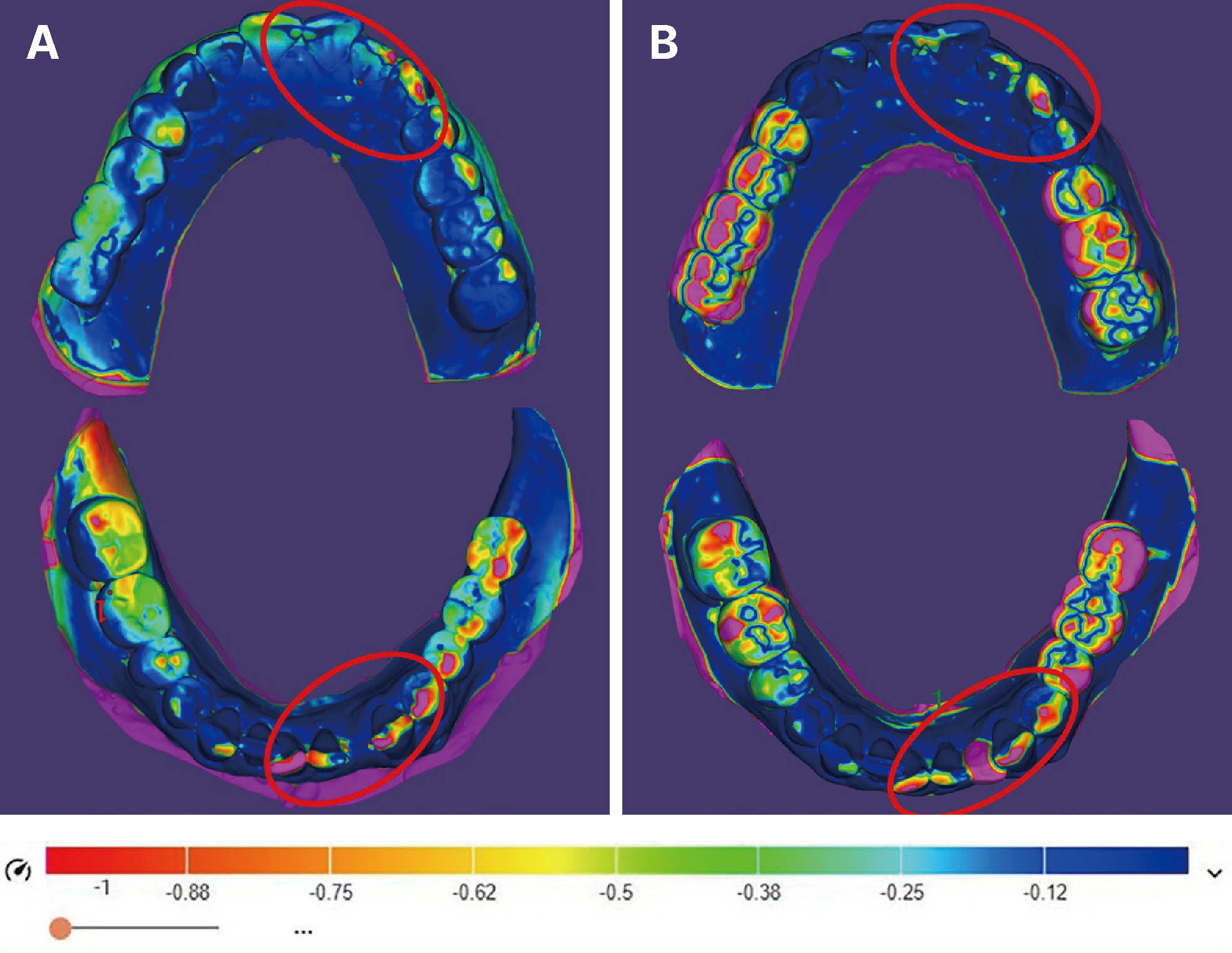
Fig. 4 Camparison of diagnostic occlusal adjustments using laboratory scan. (A) Diagnostic full mouth occlusal adjustment, (B) Diagnostic occlusal adjustment assuming posterior prosthetic reconstruction. Circle: occlusal reduction amount of residual natural teeth, unit: mm.

Fig. 5 Comparison of (A) Diagnostic full mouth occlusal adjustment and (B) Diagnostic occlusal adjustment assuming recontruction of posterior implant supported fixed prostheses.

Fig. 6 Removal of old implant supported fixed prostheses & selective occlusal adjustment of natural teeth and anterior deprogramming device. (A) Intraoral view of patient, (B) Anterior deprogramming device after selective occlusal adjustment.

Fig. 7 Comparison of (A) Diagnostic occlusal adjustment and (B) Intraoral view of patient after selective occlusal adjustment.
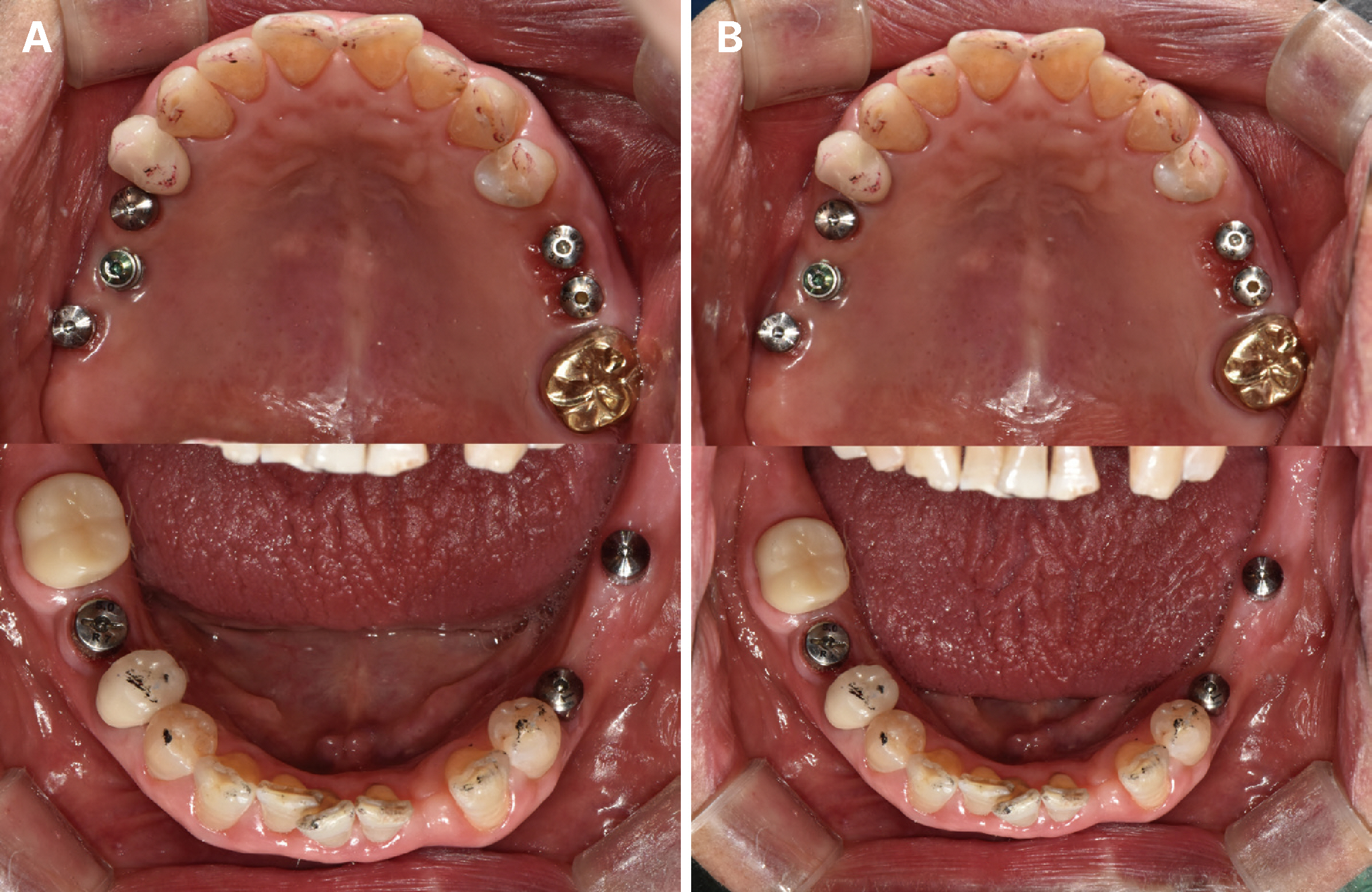
Fig. 8 Comparison of occlusal contact points after selective occlusal ajdustment. (A) Immediately after selective occlusal adjustments and (B) 3 weeks check up after selective occlusal adjustments.
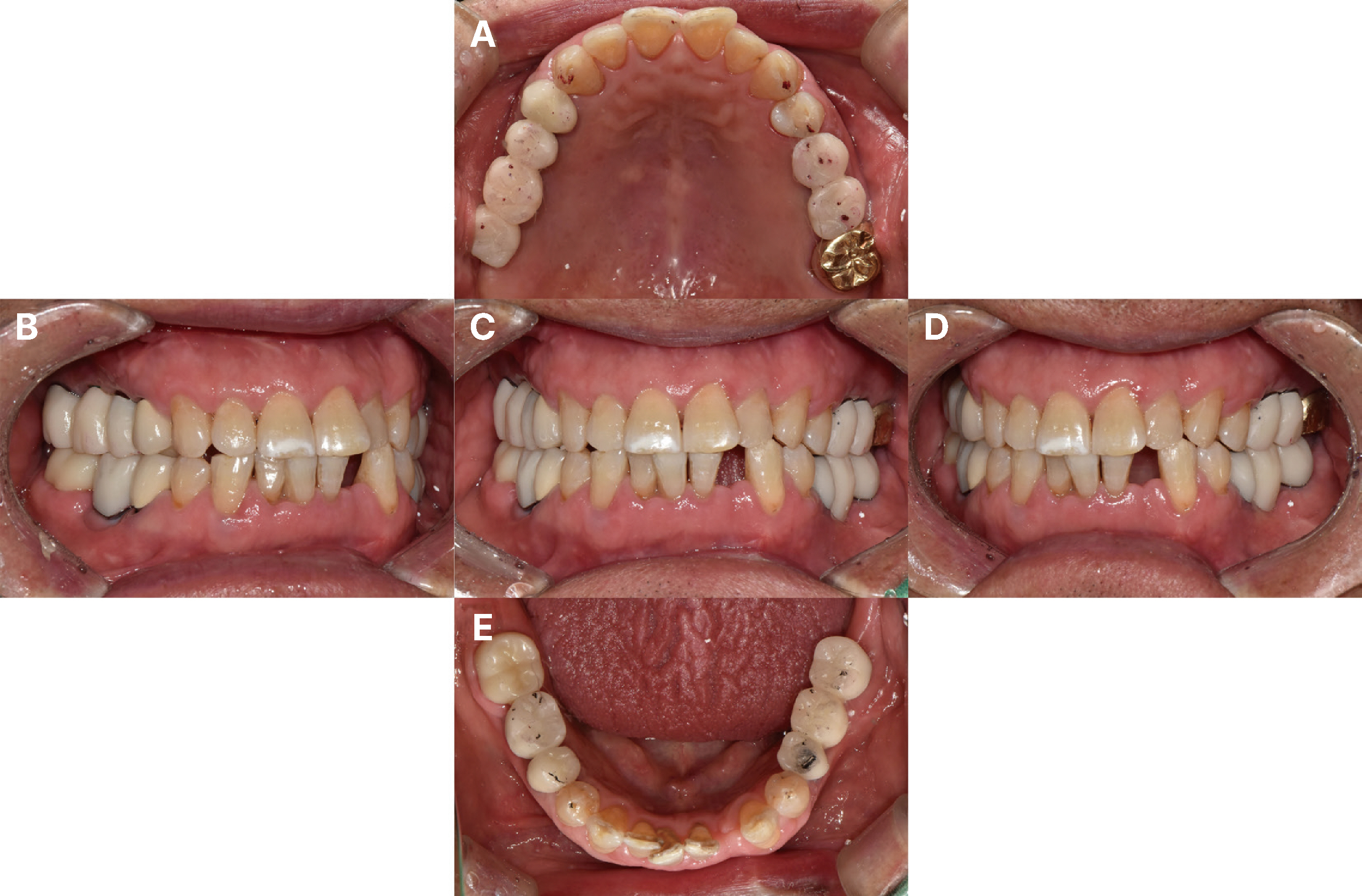
Fig. 9 Intraoral view of provisional resotrations. (A) Maxillary occlusal view, (B) Right lateral view, (C) Frontal view, (D) Left lateral view, (E) Mandibular occlusal view.
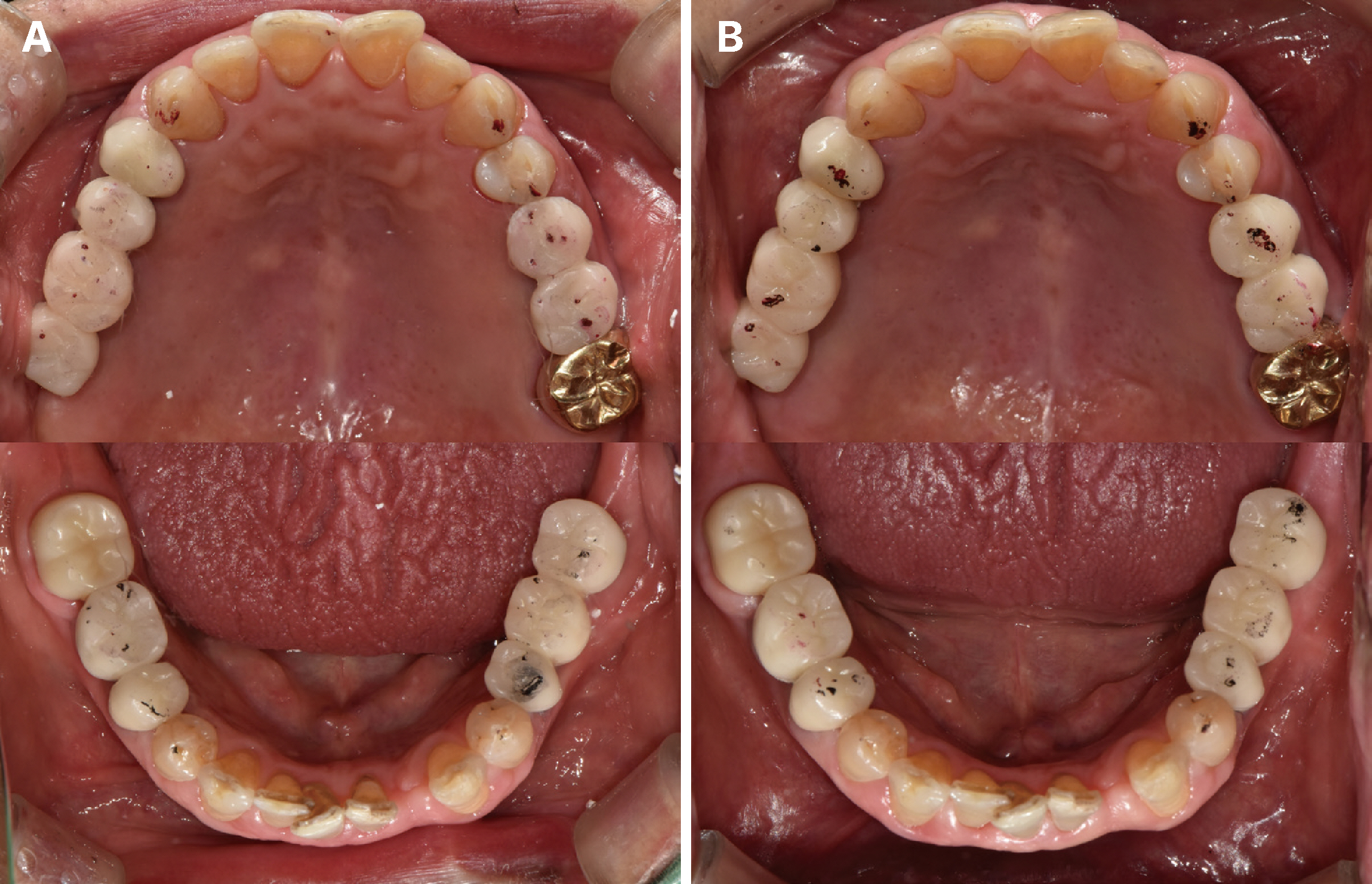
Fig. 10 Comparison of occlusal contact points of provisional restorations. (A) Immediately after setting of provisional restorations and (B) 3 weeks check up after setting of provisional restorations.

Fig. 11 Implant removal of right upper first molar site & implant placement on left lower lateral incisor site.
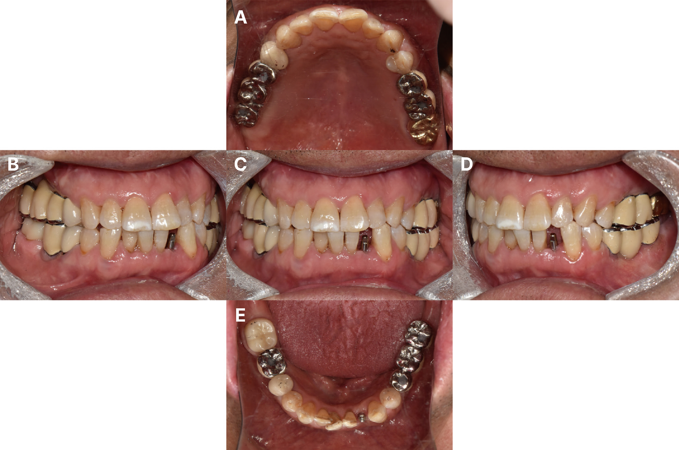
Fig. 12 Intraoral view after final setting of definitive restoration. (A) Maxillary occlusal view, (B) Right lateral view, (C) Frontal view, (D) Left lateral view, (E) Mandibular occlusal view.
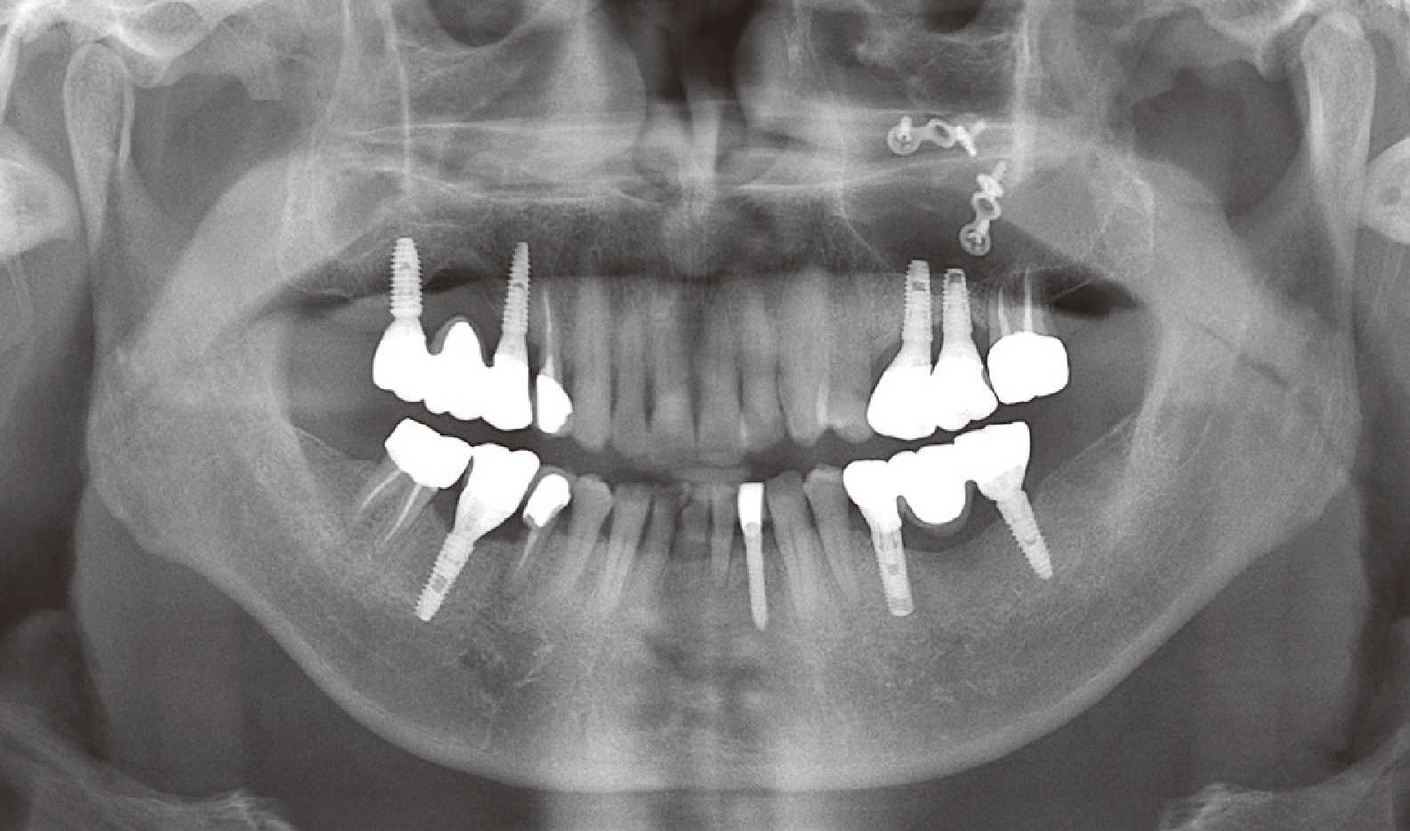
Fig. 13 Panoramic radiograph after selective occlusal adjustment & final setting of new definitive implant supported fixed prostheses.
Reference
-
References
1. Sforza C, Tartaglia GM, Lovecchio N, Ugolini A, Monteverdi R, Gianni AB, Ferrario VF. 2009; Mandibular movements at maximum mouth opening and EMG activity of masticatory and neck muscles in patients rehabilitated after a mandibular condyle fracture. J Craniomaxillofac Surg. 37:327–333. DOI: 10.1016/j.jcms.2009.01.002. PMID: 19539492.2. Korean association of oral & maxillofacial surgeons. 2013. Textbook of oral & maxillofacial surgery. 3rd ed. Dental & Medical Publishing Co.;Seoul: p. 262. p. 271.3. Pae A, Choi CH, Noh K, Kwon YD, Kim HS, Kwon KR. 2012; The prosthetic rehabilitation of a panfacial fracture patient after reduction: a clinical report. J Prosthet Dent. 108:123–8. DOI: 10.1016/S0022-3913(12)60118-8. PMID: 22867809.4. Amadi JU, Delitala F, Lieratore G, Scozzafava E, Brevi BC. 2021; Treatment decision-making for a post-traumatic malocclusion in an elderly patient: A case report. Dent Traumatol. 37:725–31. DOI: 10.1111/edt.12673. PMID: 33638228. PMCID: PMC8451768.5. Park IP, Heo SJ, Koak JY, Kim SK. 2010; Post traumatic malocclusion and its prosthetic treatment. J Adv Prosthodont. 2:88–91. DOI: 10.4047/jap.2010.2.3.88. PMID: 21165275. PMCID: PMC2994700.6. Becking AG, Zijderveld SA, Tuinzing DB. 2007; The surgical management of post-traumatic malocclusion. Clin Plast Surg. 34:e37–43. DOI: 10.1016/j.cps.2007.04.007. PMID: 17692694.7. Rozeboom A, Dubois L, Lobbezoo F, Schreurs R, Milstein D, de Lange J. 2018; Management of post-traumatic malocclusion: an alternative treatment. Oral Surg. 11:241–6. DOI: 10.1111/ors.12330.8. Dawson PE. 2006. Functional occlusion. 1st ed. Elsevier;Amsterdam: p. 59. p. 80. p. 1. DOI: 10.1016/c2012-0-07298-5.9. Kim SY, Choi YH, Kim YK. 2018; Postoperative malocclusion after maxillofacial fracture management: a retrospective case study. Maxillofac Plast Reconstr Surg. 40:27. DOI: 10.1186/s40902-018-0167-z. PMID: 30370261. PMCID: PMC6186531. PMID: 019d74cd92534abc842f63a424ee8088.10. Janson G, Crepaldi MV, Freitas KMS, de Freitas MR, Janson W. 2010; Stability of anterior open-bite treatment with occlusal adjustment. Am J Orthod Dentofacial Orthop. 138:14.e1–7. DOI: 10.1016/j.ajodo.2010.01.023. PMID: 20620828.11. Sharon E, Beyth N, Smidt A, Lipovesky-Adler M, Zilberberg N. 2019; Influence of jaw opening on occlusal vertical dimension between incisors and molars. J Prosthet Dent. 122:115–8. DOI: 10.1016/j.prosdent.2018.10.010. PMID: 30885579.
- Full Text Links
-
- Actions
-
Cited
- CITED
-
- Close
- Share
-
- Similar articles
-
- Full mouth rehabilitation of a patient with tooth wear and insufficient restorative space due to loss of posterior teeth support: a case report
- The effect of occlusal splint therapy on condylar positional changes in malocclusion patients
- Rehabilitation with orthognathic surgery and orthodontic treatment in patient with severe occlusal disharmony: A case report
- Prosthetic rehabilitation in a Class III malocclusion patient with increasing occlusal vertical dimension
- Comparison of occusal aspects in monolithic zirconia crown before and after occlusal adjustment during intraoral try-in: a case report
