Extravasation Injury of Forearm by Computed Tomography Contrast Medium
- Affiliations
-
- 1Department of Orthopedic Surgery, Daegu Fatima Hospital, Daegu, Korea. aabga@hanmail.net
- KMID: 2106660
- DOI: http://doi.org/10.4055/jkoa.2013.48.1.27
Abstract
- In recent years, there has been a noticeable increase in contrast media extravasation injury. However, definite guidelines for the treatment of the injury have not yet been established, although it causes severe complications such as compartment syndrome, skin necrosis etc. We try to introduce conservative management with a thorough review of the relevant literatures about successful treatment and functional restoration from contrast media extravasation injury without any complications.
Keyword
MeSH Terms
Figure
-
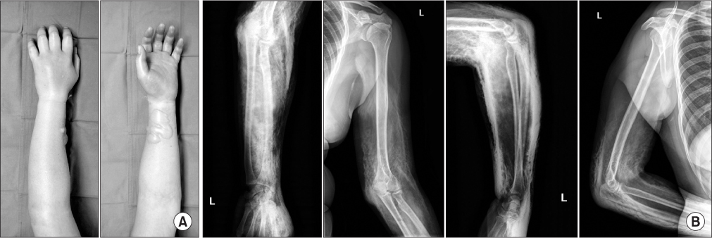
Figure 1 Clinical appearance of the upper extremity shows (A) tissue tension, global swelling, and blisters in the volar region 5 hours after the injury, and (B) radiographs of forearm and humerus 2 hours after the injury show a considerable extension of extravascular contrast.
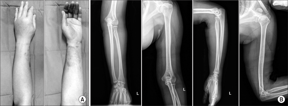
Figure 2 Clinical appearance of the upper extremity shows (A) the improvement of swelling without additional bullae formation, and (B) radiographs of the forearm and humerus show nearly contrast-absorption, except for some proximal brachium portion, 2 days after the injury.
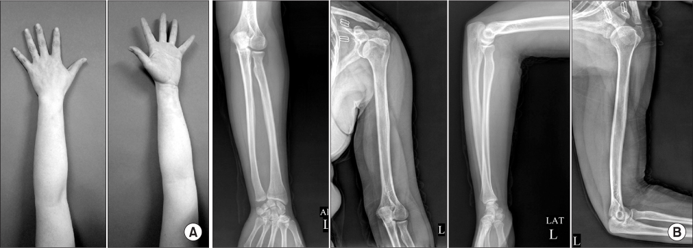
Figure 3 Clinical appearance of the upper extremity shows (A) complete remission of externals without any complications, and (B) radiographs of the forearm and humerus show no extra remaining contrast 3 months after the injury.
Reference
-
1. Cohan RH, Ellis JH, Garner WL. Extravasation of radiographic contrast material: recognition, prevention, and treatment. Radiology. 1996. 200:593–604.
Article2. Federle MP, Chang PJ, Confer S, Ozgun B. Frequency and effects of extravasation of ionic and nonionic CT contrast media during rapid bolus injection. Radiology. 1998. 206:637–640.
Article3. Loth TS, Jones DE. Extravasations of radiographic contrast material in the upper extremity. J Hand Surg Am. 1988. 13:395–398.4. Sbitany H, Koltz PF, Mays C, Girotto JA, Langstein HN. CT contrast extravasation in the upper extremity: strategies for management. Int J Surg. 2010. 8:384–386.
Article5. Schaverien MV, Evison D, McCulley SJ. Management of large volume CT contrast medium extravasation injury: technical refinement and literature review. J Plast Reconstr Aesthet Surg. 2008. 61:562–565.
Article6. Wang CL, Cohan RH, Ellis JH, Adusumilli S, Dunnick NR. Frequency, management, and outcome of extravasation of nonionic iodinated contrast medium in 69,657 intravenous injections. Radiology. 2007. 243:80–87.
Article7. Sistrom CL, Gay SB, Peffley L. Extravasation of iopamidol and iohexol during contrast-enhanced CT: report of 28 cases. Radiology. 1991. 180:707–710.
Article8. Bellin MF, Jakobsen JA, Tomassin I, et al. Contrast Media Safety Committee of the European Society of Urogenital Radiology. Contrast medium extravasation injury: guidelines for prevention and management. Eur Radiol. 2002. 12:2807–2812.
Article9. Belzunegui T, Louis CJ, Torrededia L, Oteiza J. Extravasation of radiographic contrast material and compartment syndrome in the hand: a case report. Scand J Trauma Resusc Emerg Med. 2011. 19:9.
Article10. Vandeweyer E, Heymans O, Deraemaecker R. Extravasation injuries and emergency suction as treatment. Plast Reconstr Surg. 2000. 105:109–110.
Article
- Full Text Links
-
- Actions
-
Cited
- CITED
-
- Close
- Share
-
- Similar articles
-
- Immediate surgical treatment for massive extravasation of computed tomography contrast medium in the hand: a case report
- Compartment Syndrome of Forearm Caused by Extravasation of CT Contrast Media: A Case Report
- Subcutaneous Injection Contrast Media Extravasation : 3D CT Appearance
- Compartment Syndrome of the Upper Extremity Induced by Extravasation of Contrast Media after Computed Tomography: A Case Report
- Angiographic Extravasation
