Arch Hand Microsurg.
2023 Mar;28(1):43-46. 10.12790/ahm.22.0051.
Immediate surgical treatment for massive extravasation of computed tomography contrast medium in the hand: a case report
- Affiliations
-
- 1Department of Plastic and Reconstructive Surgery, Kangbuk Samsung Hospital, Sungkyunkwan University School of Medicine, Seoul, Korea
- KMID: 2540030
- DOI: http://doi.org/10.12790/ahm.22.0051
Abstract
- There are no treatment guidelines for the extravasation of contrast medium. A 71-year-old man underwent coronary artery computed tomography. When contrast medium (iohexol) was injected through the right cephalic vein, massive leakage into the surrounding tissues occurred. A physical examination showed severe swelling of the hand and a prolonged capillary refill time. X-rays showed a massive accumulation of contrast medium in the hand. We immediately performed surgical treatment, including four skin incisions on the hand, subcutaneous dissection, and squeezing, which led to the removal of 50 mL of liquid. The immediate postoperative radiographic findings showed dramatic fading of most of the contrast medium. Four days postoperatively, radiography showed complete dissolution of all contrast medium, and a physical examination revealed full recovery of skin color and capillary refill time. We successfully managed massive extravasation of contrast medium in the hand with immediate surgical treatment. An immediate surgical approach for massive extravasation should be considered to achieve favorable outcomes.
Figure
-
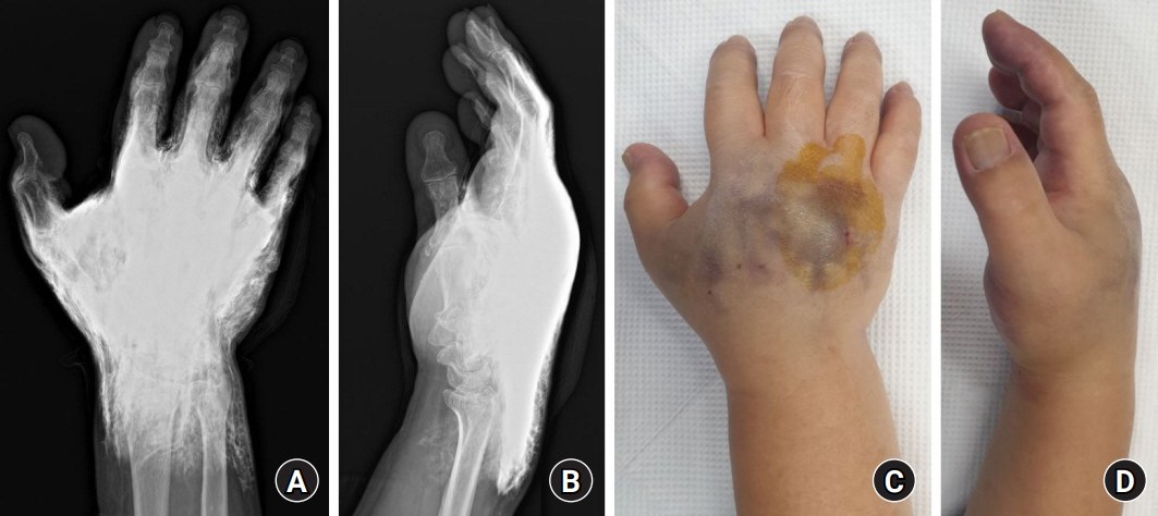
Fig. 1. Preoperative images at 1 hour after extravasation. (A, B) Preoperative X-ray shows severe swelling and marked contrast enhancement. (C, D) Preoperative image of purple discoloration on the dorsal side of the hand, indicating decreased circulation.
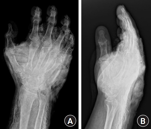
Fig. 2. Immediate postoperative X-ray images. (A, B) X-ray findings show dramatic fading of the contrast agent with decreased swelling.
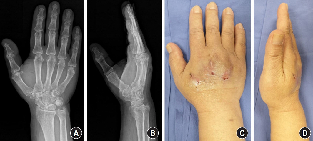
Fig. 3. Follow-up X-ray images and clinical photographs. (A, B) X-ray findings at a 5-day follow-up show no contrast agent left. (C, D) The skin color had returned to nearly normal without any necrosis at a 10-day follow-up.
Reference
-
References
1. Kaller MO, An J. Contrast Agent Toxicity [updated 2022 May 26]. In: StatPearls [Internet]. Treasure Island, FL: StatPearls Publishing;2022. Available from: https://www.ncbi.nlm.nih.gov/books/NBK537159/.2. American College of Radiology. ACR Manual on contrast media. 10th ed. Reston, VA: American College of Radiology;2017. p. 20–3.3. Federle MP, Chang PJ, Confer S, Ozgun B. Frequency and effects of extravasation of ionic and nonionic CT contrast media during rapid bolus injection. Radiology. 1998; 206:637–40.4. Heshmatzadeh Behzadi A, Farooq Z, Newhouse JH, Prince MR. MRI and CT contrast media extravasation: a systematic review. Medicine (Baltimore). 2018; 97:e0055.5. Bellin MF, Jakobsen JA, Tomassin I, et al. Contrast medium extravasation injury: guidelines for prevention and management. Eur Radiol. 2002; 12:2807–12.6. Wang CL, Cohan RH, Ellis JH, Adusumilli S, Dunnick NR. Frequency, management, and outcome of extravasation of nonionic iodinated contrast medium in 69,657 intravenous injections. Radiology. 2007; 243:80–7.7. Hadaway L. Infiltration and extravasation. Am J Nurs. 2007; 107:64–72.8. Vandeweyer E, Heymans O, Deraemaecker R. Extravasation injuries and emergency suction as treatment. Plast Reconstr Surg. 2000; 105:109–10.
- Full Text Links
-
- Actions
-
Cited
- CITED
-
- Close
- Share
-
- Similar articles
-
- Extravasation Injury of Forearm by Computed Tomography Contrast Medium
- Compartment Syndrome of Forearm Caused by Extravasation of CT Contrast Media: A Case Report
- Subcutaneous Injection Contrast Media Extravasation : 3D CT Appearance
- Computed Tomography Contrast Media Extravasation in the Upper Extremity: Clinical Features and Treatment Strategies
- Compartment Syndrome of the Upper Extremity Induced by Extravasation of Contrast Media after Computed Tomography: A Case Report
