Dural Anaplastic Large Cell Lymphoma Mimicking Meningioma: A Case Report
- Affiliations
-
- 1Department of Radiology, Seoul Paik Hospital, Inje University College of Medicine, Seoul, Korea. hongage@unitel.co.kr
- 2Department of Neurosurgery, Ilsan Paik Hospital, Inje University College of Medicine, Goyang, Korea.
- 3Department of Pathology, Seoul Paik Hospital, Inje University College of Medicine, Seoul, Korea.
- KMID: 2097998
- DOI: http://doi.org/10.3348/jksr.2014.71.4.160
Abstract
- Anaplastic large cell lymphoma (ALCL) is a rare T cell lymphoma composed of CD30-positive lymphoid cells. Most ALCLs present as nodal disease, with skin, bone, soft tissue, lung, and liver as common extranodal sites. ALCL rarely occurs in the central nervous system and is even more infrequent in the dura of the brain. We report a case of dural-based ALCL secondary to systemic disease in a 17-year-old male that mimicked meningioma on magnetic resonance imaging and angiography.
MeSH Terms
Figure
-
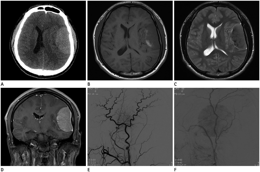
Fig. 1 Radiologic findings of dural anaplastic large cell lymphoma in a 17-year-old man. A. Precontrast axial CT shows homogenous broad dural-based semi-ovoid extra-axial mass in the left temporal lobe. B, C. On T1-weighted (B) and T2-weighted (C) MR images, a heterogeneous broad dural-based extra-axial mass in the left temporal lobe has similar signal intensity as parenchymal gray matter. D. Gadolinium-enhanced coronal T1-weighted MR images show intense and homogenous enhancement of the mass with dural-tail sign. E, F. Left external carotid artery angiogram demonstrates a prominent core vascular supply with a sunburst appearance supplied by the middle meningeal artery in the arterial phase (E) and prolonged vascular stain in the late venous phase (F).

Fig. 2 Microscopic findings of dural anaplastic large cell lymphoma in a 17-year-old man. A. Histopathologic examination under high power shows a diffuse population of large, atypical lymphoid cells with abundant cytoplasm; round and pleomorphic nuclei; and prominent nucleoli (H&E, × 200). B. Immunohistological examination under high power shows that tumor cells are immunoreactive for CD30 (immunoperoxidase, × 400).

Fig. 3 Systemic involvement of dural anaplastic large cell lymphoma in a 17-year-old man. A. Axial portal phase CT image reveals multiple enlarged and conglomerated mesenteric lymph nodes (arrows). B. Positron emission tomography image reveals multiple areas of hot uptake such as mesenteric and pelvic lymph nodes and bones of the upper and lower extremities.
Reference
-
1. Griffin JW, Thompson RW, Mitchinson MJ, De Kiewiet JC, Welland FH. Lymphomatous leptomeningitis. Am J Med. 1971; 51:200–208.2. Amaker BH, Ghatak NR, Jebraili SA, Ferreira-Gonzalez A, Kornstein MJ. Primary T-cell-rich B-cell lymphoma masquerading as a meningioma. Arch Pathol Lab Med. 2000; 124:1700–1703.3. Matmati K, Matmati N, Hannun YA, Rumboldt Z, Patel S, Lazarchick J, et al. Dural MALT lymphoma with disseminated disease. Hematol Rep. 2010; 2:e10.4. Lantos PL, Louis DN, Rosenblum MK, Kleihues P. Tumours of the nervous system. In : Graham DI, Lantos PL, editors. Greenfield's Neuropathology. 7th ed. London: Arnold;2002. vol. 2:p. 950–959.5. Miller DC, Hochberg FH, Harris NL, Gruber ML, Louis DN, Cohen H. Pathology with clinical correlations of primary central nervous system non-Hodgkin's lymphoma. The Massachusetts General Hospital experience 1958-1989. Cancer. 1994; 74:1383–1397.6. Low I, Allen J. Low-grade follicular lymphoma in the dura: rare mimic of meningioma. Neuropathology. 2006; 26:564–568.7. Villegas E, Villà S, López-Guillermo A, Petit J, Ribalta T, Graus F. Primary central nervous system lymphoma of T-cell origin: description of two cases and review of the literature. J Neurooncol. 1997; 34:157–161.8. Savage NM, Alleyne CH, Vender JR, Figueroa R, Zhang H, Samuel TA, et al. Dural-based metastatic carcinomas mimicking primary CNS neoplasia: report of 7 cases emphasizing the role of timely surgery and accurate pathologic evaluation. Int J Clin Exp Pathol. 2011; 4:530–540.
- Full Text Links
-
- Actions
-
Cited
- CITED
-
- Close
- Share
-
- Similar articles
-
- A Subcortical Anaplastic Meningioma
- Dermatofibroma in Patient with Relapsing Primary Cutaneous Anaplastic Large Cell Lymphoma
- Skeletal Muscle Lymphoma Mimicking Abscess
- Anaplastic Large Cell Lymphoma Mimicking a Muscle Abscess: A Case Report
- A Case of ALK-Negative Systemic Anaplastic Large Cell Lymphoma
