Tissue reactions to suture materials in the oral mucosa of beagle dogs
- Affiliations
-
- 1Luden Dental Clinic, Seoul, Korea.
- 2Department of Periodontology, Kyung Hee University School of Dentistry, Seoul, Korea. chungjh@khu.ac.kr
- 3Institute of Oral Biology, Kyung Hee University School of Dentistry, Seoul, Korea.
- KMID: 2094718
- DOI: http://doi.org/10.5051/jpis.2011.41.4.185
Abstract
- PURPOSE
The objective of this study was to compare and evaluate the inflammatory responses of three widely used suture materials in the keratinized gingiva and buccal mucosa of beagle dogs.
METHODS
Silk, polyglycolic acid, and nylon sutures were placed within the mandibular keratinized gingiva and maxillary buccal mucosa of four male beagle dogs. Biopsies were taken 3, 7, and 14 days after suturing. Specimens were prepared with hematoxylin-eosin stain for evaluation under a light microscope.
RESULTS
The suture materials placed in the oral mucosa elicited more inflammatory reactions than did those placed in the keratinized gingiva. The multifilament suture materials caused more inflammatory tissue reactions than did the monofilament suture materials in the oral mucosa.
CONCLUSIONS
If oral hygiene is well maintained and suture materials are placed in the keratinized gingiva, silk, nylon, and polyglycolic acid are considered to be proper suture materials for oral surgery. However, it is advisable to use monofilament suture materials if the suture site is within the oral mucosa.
Keyword
MeSH Terms
Figure
-
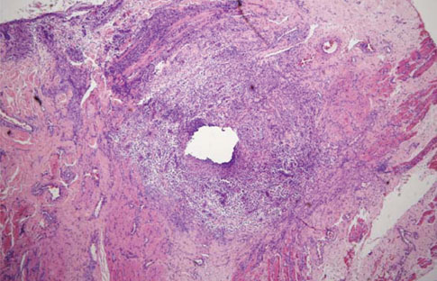
Figure 1 Silk suture at seven days in buccal oral mucosa. The suture loop is surrounded by dense inflammatory cell infiltrate and a zone of inflammatory reaction (H&E staining, original magnification ×40).
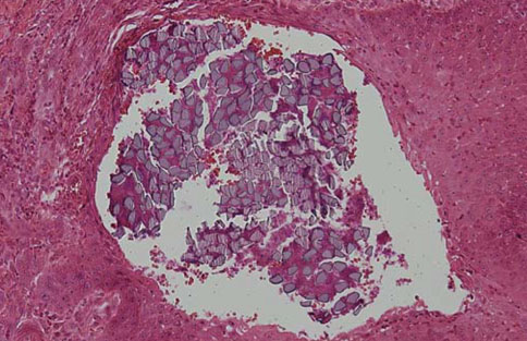
Figure 2 Silk suture at 14 days in keratinized gingiva. Bacterial plaque and inflammatory cell infiltration in the suture filaments is observed. Epithelial invagination is well advanced, and the suture material is lined by epithelium (H&E staining, original magnification ×200).

Figure 3 Silk suture at 14 days in buccal oral mucosa. Numerous polymorphonuclear leucocytes and lymphocytes are evident. Epithelial cells are observed around the suture material (H&E staining, original magnification ×400).
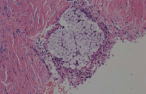
Figure 4 Polyglycolic acid suture at three days in keratinized gingiva. A few lymphocytes and moderate polymorphonuclear leucocyte infiltration are observed around the suture material. Very few lymphocytes and macrophages are observed in the interstitium between the collagen bundles (H&E staining, original magnification ×200).
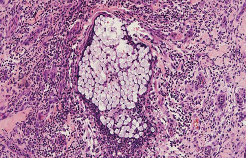
Figure 5 Polyglycolic acid suture at seven days in buccal oral mucosa. Inflammatory cells are present within the suture, as well as in the adjacent submucosal area. The suture material is still intact (H&E staining, original magnification ×200).
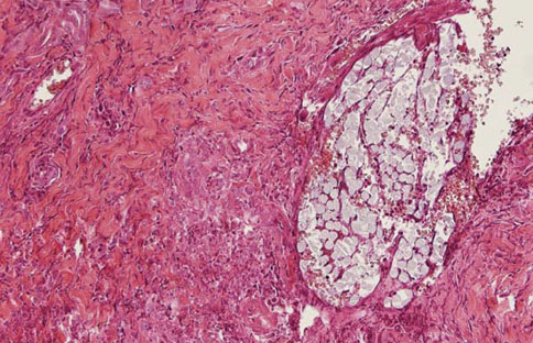
Figure 6 Polyglycolic acid suture at 14 days in keratinized gingiva. There is migration of monocytes into the interstices. The perisutural granulation tissue contains a few lymphocytes and macrophages, as well as elongated collagen fibers (H&E staining, original magnification ×200).
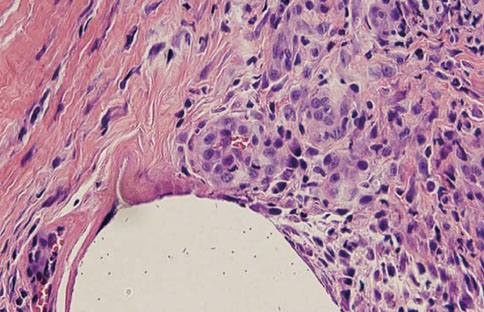
Figure 7 Nylon suture at seven days in keratinized gingiva. The perisutural tissue contains predominantly polymorphonuclear leucocytes and lymphocytes. Newly formed collagen fibrils can be observed (H&E staining, original magnification ×400).
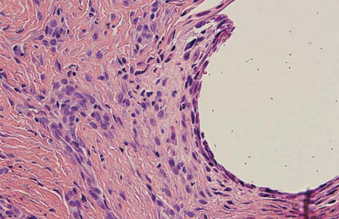
Figure 8 Nylon suture at 14 days in keratinized gingiva. The microscopic findings around the suture reveal some neutrophils and lymphocyte infiltration. Perisutural granulation tissue containing many capillaries and collagen fibrils is visible. Moderate inflammatory reaction can be observed (H&E staining, original magnification ×400).
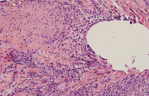
Figure 9 Nylon suture at 14 days in buccal oral mucosa. Significant inflammatory cell infiltration--polymorphonuclear leucocytes, macrophages, and lymphocytes--is observed along the suture track (H&E staining, original magnification ×200).
Reference
-
1. Shaw RJ, Negus TW, Mellor TK. A prospective clinical evaluation of the longevity of resorbable sutures in oral mucosa. Br J Oral Maxillofac Surg. 1996. 34:252–254.
Article2. Swanson NA, Tromovitch TA. Suture materials, 1980s: properties, uses, and abuses. Int J Dermatol. 1982. 21:373–378.
Article3. Cohen ES. Atlas of cosmetic and reconstructive periodontal surgery. 2007. 3rd ed. Hamilton: BC Decker.4. Grigg TR, Liewehr FR, Patton WR, Buxton TB, McPherson JC. Effect of the wicking behavior of multifilament sutures. J Endod. 2004. 30:649–652.
Article5. Ivanoff CJ, Widmark G. Nonresorbable versus resorbable sutures in oral implant surgery: a prospective clinical study. Clin Implant Dent Relat Res. 2001. 3:57–60.
Article6. Craig PH, Williams JA, Davis KW, Magoun AD, Levy AJ, Bogdansky S, et al. A biologic comparison of polyglactin 910 and polyglycolic acid synthetic absorbable sutures. Surg Gynecol Obstet. 1975. 141:1–10.7. Silverstein LH. Principles of dental suturing: the complete guide to surgical closure. 1999. New Jersey: Montage Media Co..8. Selvig KA, Biagiotti GR, Leknes KN, Wikesjo UM. Oral tissue reactions to suture materials. Int J Periodontics Restorative Dent. 1998. 18:474–487.9. Sanz L, Smith S. Mechanisms of wound healing, suture material, and wound closure, strategies in gynecologic surgery. 1986. New York: Springer.10. Yaltirik M, Dedeoglu K, Bilgic B, Koray M, Ersev H, Issever H, et al. Comparison of four different suture materials in soft tissues of rats. Oral Dis. 2003. 9:284–286.
Article11. Leknes KN, Røynstrand IT, Selvig KA. Human gingival tissue reactions to silk and expanded polytetrafluoroethylene sutures. J Periodontol. 2005. 76:34–42.
Article12. Lilly GE, Armstrong JH, Salem JE, Cutcher JL. Reaction of oral tissues to suture materials. II. Oral Surg Oral Med Oral Pathol. 1968. 26:592–599.13. Rothenburger S, Spangler D, Bhende S, Burkley D. In vitro antimicrobial evaluation of Coated VICRYL* Plus Antibacterial Suture (coated polyglactin 910 with triclosan) using zone of inhibition assays. Surg Infect (Larchmt). 2002. 3:Suppl 1. S79–S87.
Article14. Gabrielli F, Potenza C, Puddu P, Sera F, Masini C, Abeni D. Suture materials and other factors associated with tissue reactivity, infection, and wound dehiscence among plastic surgery outpatients. Plast Reconstr Surg. 2001. 107:38–45.
Article15. Castelli WA, Nasjleti CF, Diaz-Perez R, Caffesse RG. Cheek mucosa response to silk, cotton, and nylon suture materials. Oral Surg Oral Med Oral Pathol. 1978. 45:186–189.
Article16. Racey GL, Wallace WR, Cavalaris CJ, Marguard JV. Comparison of a polyglycolic-polylactic acid suture to black silk and plain catgut in human oral tissues. J Oral Surg. 1978. 36:766–770.17. Blomstedt B, Osterberg B, Bergstrand A. Suture material and bacterial transport. An experimental study. Acta Chir Scand. 1977. 143:71–73.18. King RC, Crawford JJ, Small EW. Bacteremia following intraoral suture removal. Oral Surg Oral Med Oral Pathol. 1988. 65:23–28.
Article19. Blumenthal NM. A clinical comparison of collagen membranes with e-PTFE membranes in the treatment of human mandibular buccal class II furcation defects. J Periodontol. 1993. 64:925–933.
Article20. Cardaropoli G, Araújo M, Lindhe J. Dynamics of bone tissue formation in tooth extraction sites. An experimental study in dogs. J Clin Periodontol. 2003. 30:809–818.21. Romanos GE, Schröter-Kermani C, Weingart D, Strub JR. Health human periodontal versus peri-implant gingival tissues: an immunohistochemical differentiation of the extracellular matrix. Int J Oral Maxillofac Implants. 1995. 10:750–758.22. Lilly GE. Reaction of oral tissues to suture materials. Oral Surg Oral Med Oral Pathol. 1968. 26:128–133.
Article23. Sortino F, Lombardo C, Sciacca A. Silk and polyglycolic acid in oral surgery: a comparative study. Oral Surg Oral Med Oral Pathol Oral Radiol Endod. 2008. 105:e15–e18.
Article24. Gabel EA, Jimenez GP, Eaglstein WH, Kerdel FA, Falanga V. Performance comparison of nylon and an absorbable suture material (Polyglactin 910) in the closure of punch biopsy sites. Dermatol Surg. 2000. 26:750–752.
Article25. Leknes KN, Selvig KA, Bøe OE, Wikesjö UM. Tissue reactions to sutures in the presence and absence of anti-infective therapy. J Clin Periodontol. 2005. 32:130–138.
Article
- Full Text Links
-
- Actions
-
Cited
- CITED
-
- Close
- Share
-
- Similar articles
-
- An experimental study on gross reactions of surrounding maxillary sutures to the widening of midpalatal suture in the dog
- Molecular identification of Mycoplasma cynos from laboratory beagle dogs with respiratory disease
- Effects of sodium butyrate-containing calcium sulfate bone graft on oral mucosa and bone tissue
- A comparative experimental study on gross reactions of surounding maxillary sutures to the widening of midpalatal suture in young and adult dog
- Comparative Study of Tissue Response of Various Suture Materials in Rats
