Korean J Hematol.
2007 Jun;42(2):172-175. 10.5045/kjh.2007.42.2.172.
A Case of Unspecified Mature T-cell Leukemia with Clover-shaped, Multi-lobated Nuclei
- Affiliations
-
- 1Department of Laboratory Medicine, Samsung Medical Center, Sungkyunkwan University School of Medicine, Seoul, Korea. sunnyhk@smc.samsung.co.kr
- KMID: 2083517
- DOI: http://doi.org/10.5045/kjh.2007.42.2.172
Abstract
- Mature T-cell leukemias are a group of neoplasms derived from mature or post-thymic T-cells, and a number of distinctive disease entities have been defined in the World Health Organization (WHO) classification. Here we report a 54-year-old female patient with multi-lobated atypical cells expressing the classic T-cell antigens involving multiple lymph nodes, peripheral blood, and bone marrow. The clinical, laboratory, and pathologic features of her disease did not fit into any of the entities in the WHO Classification. There was no evidence of rapidly rising lymphocyte counts, TCL1 expression, eosinophilia, erythroderma, Sezary cells, autoimmune phenomena, cytotoxic granules, nor evidence of HTLV-1 infection, and thus, T-cell prolymphocytic leukemia, Sezary syndrome, T-cell granular lymphocytic leukemia, and adult T-cell leukemia/lymphoma were all ruled out. This case suggests that further characterization and definition of the "unclassifiable" cases of mature T-cell neoplasm is needed to better understand the group of disorders.
MeSH Terms
Figure
-
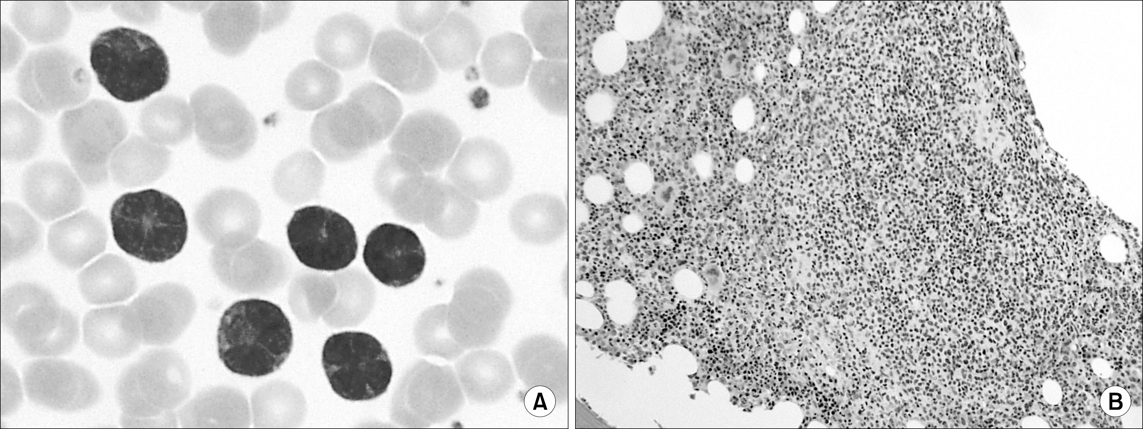
Fig. 1 (A) Medium-sized atypical lymphocytes with clover-shaped multi-lobated nuclei on marrow aspirate (Wright- Giemsa stain, ×1,000). (B) Nodular infiltration of the atypical neoplastic cells on marrow biopsy (Hematoxylin-Eosin stain, ×100).
Reference
-
1). Ravandi F., Kantarjian H., Jones D., Dearden C., Keating M., O'Brien S. Mature T-cell leukemias. Cancer. 2005. 104:1808–18.
Article2). Jaffe ES HN., Stein H, et al. World Health Organization classification of tumours: pathology and genetics of tumours of haematopoietic and lymphoid tissues. 1st ed.Lyon, France: IARC Press;2001. p. 190–203.3). Herling M., Khoury JD., Washington LT., Duvic M., Keating MJ., Jones D. A systematic approach to diagnosis of mature T-cell leukemias reveals heterogeneity among WHO categories. Blood. 2004. 104:328–35.
Article4). Matutes E. T-cell prolymphocytic leukemia. Cancer Control. 1998. 5:19–24.
Article5). Matutes E., Garcia Talavera J., O'Brien M., Catovsky D. The morphological spectrum of T-prolympho-cytic leukaemia. Br J Haematol. 1986. 64:111–24.
Article6). Foss F. Mycosis fungoides and the Sézary syndrome. Curr Opin Oncol. 2004. 16:421–8.
Article7). Shimoyama M. Diagnostic criteria and classification of clinical subtypes of adult T-cell leukaemia-lymphoma. A report from the Lymphoma Study Group (1984∼87). Br J Haematol. 1991. 79:428–37.8). Miyamoto K., Kagami Y., Shimoyama M, et al. A unique T-cell line derived from an HTLV-1-negative adult T-cell leukemia patient. Jpn J Cancer Res. 1987. 78:1031–5.9). Kirito K., Shindou H., Chiba N., Tobinai K., Shimoyama M., Kinoshita T. HTLV-I negative adult T cell leukemia; a case report of acute type. Rinsho Ketsue-ki. 1993. 34:1550–5.
