Primary malignant melanoma of the uterine cervix: A case report
- Affiliations
-
- 1Department Obstetrics and Gynecology, Inha University College of Medicine, Incheon, Korea. sohwang@inha.ac.kr
- 2Department Pathology, Inha University College of Medicine, Incheon, Korea.
- KMID: 2078152
- DOI: http://doi.org/10.5468/KJOG.2012.55.5.343
Abstract
- After the discovery of melanocytes in the cervix in 1959, it was recognized that primary malignant melanoma of the cervix exists as a separate entity. A 74-year-old woman visited hospital for vaginal bleeding from a black colored cervical mass. The pathology of cervical punch biopsy showed a malignant melanoma with positive immnunohistochemical stainings for S100 protein and HMB-45 antibody. Abdominal radical hysterectomy with pelvic and paraaortic lymphadenectomy was performed. The final pathology was a malignant melanoma of the cervix with metastases for both external iliac lymph nodes and tumor involvement in the margin of vaginal resection. She received cisplatin based concurrent chemoradiotherapy postoperatively. But 6 months later, she received another chemotherapy with dacarbazine and cisplatin for recurrence. We report a case of a 74-year-old patient with a malignant melanoma of the uterine cervix with a brief review.
Keyword
MeSH Terms
Figure
-
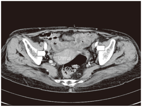
Fig. 1 Computed tomography findings of preoperation; upper margin of mild enhancing cervical mass. The multiple small lymph nodes in both external iliac chains were enlarged.
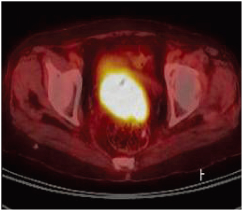
Fig. 2 Positron emission tomography findings of abnormal fluoro-D-glucose uptake in whole cervix mass.
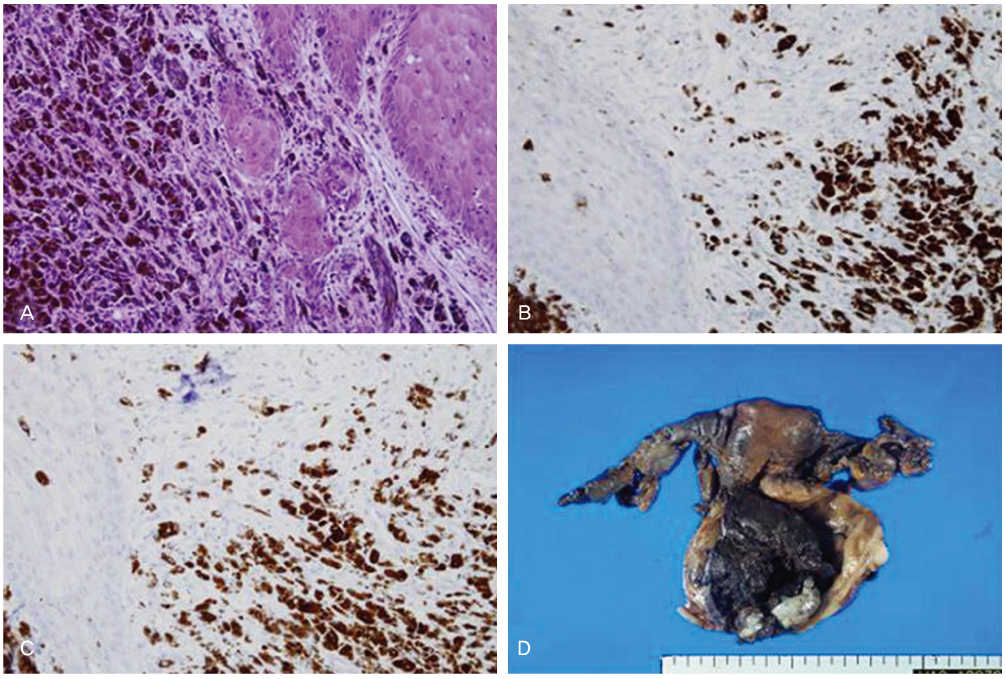
Fig. 3 Histologic findings are showing proliferation of malignant melanin-producing melanocytes (A). Most of the atypical cells are positive for HMB-45 (B) and S100 (C) (A: H&E, ×20; B: Immunohistochemical stain, ×20; C: Immunohistochemical stain for S100 protein after melanin bleaching, ×20). The uterus was enlarged with a huge dark brown ulcerofungating tumor arising in the exocervix and endocervix, extending to the vagina with near full-thickness invasion of the cervical wall (D).
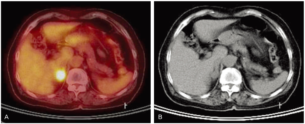
Fig. 4 About 6 months after radical hysterectomy, newly appeared multiple well-defined various size pulmonary nodule in right lung and left lower lobe that suspected pulmonary metastasis. Computed tomography findings of round shape well-defined 0.6 cm and 0.4 cm sized pulmonary nodules in right lower lobe (allow).
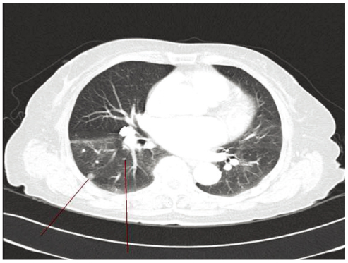
Fig. 5 Positron emission tomography-computed tomography imaging. Abnormal FDG uptake in newly appeared 2 cm sized right adrenal gland nodule 6 month after radical hysterectomy.
Reference
-
1. Cid JM. Melanoid pigmentation of the endocervix: a neurogenic visceral argument. Ann Anat Pathol (Paris). 1959. 4:617–628.2. Podczaski E, Abt A, Kaminski P, Larson J, Sorosky J, DeGeest K, et al. A patient with multiple, malignant melanomas of the lower genital tract. Gynecol Oncol. 1990. 37:422–426.3. Gupta R, Singh S, Mandal AK. Primary malignant melanoma of cervix - a case report. Indian J Cancer. 2005. 42:201–204.4. Ma SQ, Bai CM, Zhong S, Yu XH, Lang JH. Clinical analysis of primary malignant melanoma of the cervix. Chin Med Sci J. 2005. 20:257–260.5. Mousavi AS, Fakor F, Nazari Z, Ghaemmaghami F, Hashemi FA, Jamali M. Primary malignant melanoma of the uterine cervix: case report and review of the literature. J Low Genit Tract Dis. 2006. 10:258–263.6. DeMatos P, Tyler D, Seigler HF. Mucosal melanoma of the female genitalia: a clinicopathologic study of forty-three cases at Duke University Medical Center. Surgery. 1998. 124:38–48.7. Clark KC, Butz WR, Hapke MR. Primary malignant melanoma of the uterine cervix: case report with world literature review. Int J Gynecol Pathol. 1999. 18:265–273.8. Mordel N, Mor-Yosef S, Ben-Baruch N, Anteby SO. Malignant melanoma of the uterine cervix: case report and review of the literature. Gynecol Oncol. 1989. 32:375–380.9. Deshpande AH, Munshi MM. Primary malignant melanoma of the uterine cervix: report of a case diagnosed by cervical scrape cytology and review of the literature. Diagn Cytopathol. 2001. 25:108–111.10. Norris HJ, Taylor HB. Melanomas of the vagina. Am J Clin Pathol. 1966. 46:420–426.11. Hytiroglou P, Domingo J. Development of melanosis of uterine cervix after cryotherapy for epithelial dysplasia. A case report and brief review of the literature on pigmented lesions of the cervix. Am J Clin Pathol. 1990. 93:802–805.12. Kristiansen SB, Anderson R, Cohen DM. Primary malignant melanoma of the cervix and review of the literature. Gynecol Oncol. 1992. 47:398–403.13. Cantuaria G, Angioli R, Nahmias J, Estape R, Penalver M. Primary malignant melanoma of the uterine cervix: case report and review of the literature. Gynecol Oncol. 1999. 75:170–174.14. Herbert SH, Solin LJ, Rate WR, Schultz DJ, Hanks GE. The effect of palliative radiation therapy on epidural compression due to metastatic malignant melanoma. Cancer. 1991. 67:2472–2476.
- Full Text Links
-
- Actions
-
Cited
- CITED
-
- Close
- Share
-
- Similar articles
-
- A Case report of Primary Malignant Melanoma of the Uterine Cervix
- A Case of Primary Malignant Melanoma of Uterine Cervix
- Primary amelanotic melanoma of the cervix: case report with review of literature
- A case of synchronous multiple primary malignant neoplasm of the uterine cervix and endometrium
- A Case of Primary Malignant Lymphoma of the Uterine Cervix
