Effect of Increasing Diffusion Gradient Direction Number on Diffusion Tensor Imaging Fiber Tracking in the Human Brain
- Affiliations
-
- 1School of Optical-Electrical and Computer Engineering, Shanghai Medical Instrument College, University of Shanghai for Science and Technology, Shanghai 200093, China. slzhuangx@yahoo.com
- 2Department of Radiology, Huashan Hospital, Fudan University, Shanghai 200040, China.
- KMID: 2070185
- DOI: http://doi.org/10.3348/kjr.2015.16.2.410
Abstract
OBJECTIVE
To assess the effects of varying the number of diffusion gradient directions (NDGDs) on diffusion tensor fiber tracking (FT) in human brain white matter using tract characteristics.
MATERIALS AND METHODS
Twelve normal volunteers underwent diffusion tensor imaging (DTI) scanning with NDGDs of 6, 11, 15, 21, and 31 orientations. Three fiber tract groups, including the splenium of the corpus callosum (CC), the entire CC, and the full brain tract, were reconstructed by deterministic DTI-FT. Tract architecture was first qualitatively evaluated by visual observation. Six quantitative tract characteristics, including the number of fibers (NF), average length (AL), fractional anisotropy (FA), relative anisotropy (RA), mean diffusivity (MD), and volume ratio (VR) were measured for the splenium of the CC at the tract branch level, for the entire CC at tract level, and for the full brain tract at the whole brain level. Visual results and those of NF, AL, FA, RA, MD, and VR were compared among the five different NDGDs.
RESULTS
The DTI-FT with NDGD of 11, 15, 21, and 31 orientations gave better tracking results compared with NDGD of 6 after the visual evaluation. NF, FA, RA, MD, and VR values with NDGD of six were significantly greater (smallest p = 0.001 to largest p = 0.042) than those with four other NDGDs (11, 15, 21, or 31 orientations), whereas AL measured with NDGD of six was significantly smaller (smallest p = 0.001 to largest p = 0.041) than with four other NDGDs (11, 15, 21, or 31 orientations). No significant differences were observed in the results among the four NDGD groups of 11, 15, 21, and 31 directions (smallest p = 0.059 to largest p = 1.000).
CONCLUSION
The main fiber tracts were detected with NDGD of six orientations; however, the use of larger NDGD (> or = 11 orientations) could provide improved tract characteristics at the expense of longer scanning time.
Keyword
MeSH Terms
Figure
-

Fig. 1 Comparison of splenium of corpus callosum using diffusion tensor imaging fiber tracking with five diffusion gradient directions (NDGDs). A. NDGD of 6. B. NDGD of 11. C. NDGD of 15. D. NDGD of 21. E. NDGD of 31. As described in this paper, higher NDGD (11, 15, 21, and 31 orientations) (green arrows in B-E) detected longer fibers that extended into bilateral temporal lobes. Here, green arrows are used to emphasize demonstration of increasing NDGD on fiber tracking at higher NDGDs.
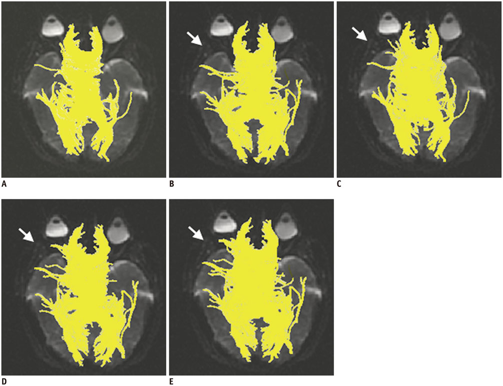
Fig. 2 Comparison of corpus callosum using diffusion tensor imaging fiber tracking with five diffusion gradient directions (NDGDs). A. NDGD of 6. B. NDGD of 11. C. NDGD of 15. D. NDGD of 21. E. NDGD of 31. As described in this paper, more branches of corpus callosum were propagated with increasing NDGD, and there were no main differences at NDGDs of 11, 15, 21, and 31, as shown in B-E by white arrows. Here, white arrows are implemented to point out variation of increasing NDGD on fiber tracking at higher NDGDs.

Fig. 3 Comparison of full brain tract using diffusion tensor imaging fiber tracking with five diffusion gradient directions (NDGDs). A. NDGD of 6. B. NDGD of 11. C. NDGD of 15. D. NDGD of 21. E. NDGD of 31. As described in this paper, more tracts were extended to brainstem and cerebellum with high fractional anisotropy (FA) values (labeled by yellow arrows) at five higher NDGDs. Some low FA value tracts in parietal lobe showed different configurations; one such difference is indicated by red arrows. Used arrows are applied to distinguish differences of fiber tracking with increasing NDGDs.
Cited by 1 articles
-
Diffusion Tensor-Derived Properties of Benign Oligemia, True “at Risk” Penumbra, and Infarct Core during the First Three Hours of Stroke Onset: A Rat Model
Fang-Ying Chiu, Duen-Pang Kuo, Yung-Chieh Chen, Yu-Chieh Kao, Hsiao-Wen Chung, Cheng-Yu Chen
Korean J Radiol. 2018;19(6):1161-1171. doi: 10.3348/kjr.2018.19.6.1161.
Reference
-
1. Pierpaoli C, Jezzard P, Basser PJ, Barnett A, Di Chiro G. Diffusion tensor MR imaging of the human brain. Radiology. 1996; 201:637–648.2. Mori S, van Zijl PC. Fiber tracking: principles and strategies - a technical review. NMR Biomed. 2002; 15:468–480.3. Descoteaux M, Deriche R, Knösche TR, Anwander A. Deterministic and probabilistic tractography based on complex fibre orientation distributions. IEEE Trans Med Imaging. 2009; 28:269–286.4. Berman JI, Mukherjee P, Partridge SC, Miller SP, Ferriero DM, Barkovich AJ, et al. Quantitative diffusion tensor MRI fiber tractography of sensorimotor white matter development in premature infants. Neuroimage. 2005; 27:862–871.5. Mukherjee P, Chung SW, Berman JI, Hess CP, Henry RG. Diffusion tensor MR imaging and fiber tractography: technical considerations. AJNR Am J Neuroradiol. 2008; 29:843–852.6. Anwander A, Tittgemeyer M, von Cramon DY, Friederici AD, Knösche TR. Connectivity-Based Parcellation of Broca's Area. Cereb Cortex. 2007; 17:816–825.7. Fillard P, Pennec X, Arsigny V, Ayache N. Clinical DT-MRI estimation, smoothing, and fiber tracking with log-Euclidean metrics. IEEE Trans Med Imaging. 2007; 26:1472–1482.8. Nimsky C, Ganslandt O, Merhof D, Sorensen AG, Fahlbusch R. Intraoperative visualization of the pyramidal tract by diffusion-tensor-imaging-based fiber tracking. Neuroimage. 2006; 30:1219–1229.9. Jeurissen B, Leemans A, Jones DK, Tournier JD, Sijbers J. Probabilistic fiber tracking using the residual bootstrap with constrained spherical deconvolution. Hum Brain Mapp. 2011; 32:461–479.10. Makki MI, Govindan RM, Wilson BJ, Behen ME, Chugani HT. Altered fronto-striato-thalamic connectivity in children with Tourette syndrome assessed with diffusion tensor MRI and probabilistic fiber tracking. J Child Neurol. 2009; 24:669–678.11. Roosendaal SD, Geurts JJ, Vrenken H, Hulst HE, Cover KS, Castelijns JA, et al. Regional DTI differences in multiple sclerosis patients. Neuroimage. 2009; 44:1397–1403.12. Torgerson CM, Irimia A, Leow AD, Bartzokis G, Moody TD, Jennings RG, et al. DTI tractography and white matter fiber tract characteristics in euthymic bipolar I patients and healthy control subjects. Brain Imaging Behav. 2013; 7:129–139.13. Chahboune H, Mishra AM, DeSalvo MN, Staib LH, Purcaro M, Scheinost D, et al. DTI abnormalities in anterior corpus callosum of rats with spike-wave epilepsy. Neuroimage. 2009; 47:459–466.14. Kantarci K, Avula R, Senjem ML, Samikoglu AR, Zhang B, Weigand SD, et al. Dementia with Lewy bodies and Alzheimer disease: neurodegenerative patterns characterized by DTI. Neurology. 2010; 74:1814–1821.15. Lenglet C, Campbell JS, Descoteaux M, Haro G, Savadjiev P, Wassermann D, et al. Mathematical methods for diffusion MRI processing. Neuroimage. 2009; 45:1 Suppl. S111–S122.16. Ni H, Kavcic V, Zhu T, Ekholm S, Zhong J. Effects of number of diffusion gradient directions on derived diffusion tensor imaging indices in human brain. AJNR Am J Neuroradiol. 2006; 27:1776–1781.17. Bammer R, Holdsworth SJ, Veldhuis WB, Skare ST. New methods in diffusion-weighted and diffusion tensor imaging. Magn Reson Imaging Clin N Am. 2009; 17:175–204.18. Papadakis NG, Murrills CD, Hall LD, Huang CL, Adrian Carpenter T. Minimal gradient encoding for robust estimation of diffusion anisotropy. Magn Reson Imaging. 2000; 18:671–679.19. Zhan L, Leow AD, Jahanshad N, Chiang MC, Barysheva M, Lee AD, et al. How does angular resolution affect diffusion imaging measures? Neuroimage. 2010; 49:1357–1371.20. Lebel C, Benner T, Beaulieu C. Six is enough? Comparison of diffusion parameters measured using six or more diffusion-encoding gradient directions with deterministic tractography. Magn Reson Med. 2012; 68:474–483.21. Heiervang E, Behrens TE, Mackay CE, Robson MD, Johansen-Berg H. Between session reproducibility and between subject variability of diffusion MR and tractography measures. Neuroimage. 2006; 33:867–877.22. Roberts TP, Liu F, Kassner A, Mori S, Guha A. Fiber density index correlates with reduced fractional anisotropy in white matter of patients with glioblastoma. AJNR Am J Neuroradiol. 2005; 26:2183–2186.23. Jiang H, van Zijl PC, Kim J, Pearlson GD, Mori S. DtiStudio: resource program for diffusion tensor computation and fiber bundle tracking. Comput Methods Programs Biomed. 2006; 81:106–116.24. Le Bihan D, Mangin JF, Poupon C, Clark CA, Pappata S, Molko N, et al. Diffusion tensor imaging: concepts and applications. J Magn Reson Imaging. 2001; 13:534–546.25. Jellison BJ, Field AS, Medow J, Lazar M, Salamat MS, Alexander AL. Diffusion tensor imaging of cerebral white matter: a pictorial review of physics, fiber tract anatomy, and tumor imaging patterns. AJNR Am J Neuroradiol. 2004; 25:356–369.26. Bello L, Gambini A, Castellano A, Carrabba G, Acerbi F, Fava E, et al. Motor and language DTI Fiber Tracking combined with intraoperative subcortical mapping for surgical removal of gliomas. Neuroimage. 2008; 39:369–382.27. Yao X, Wang M, Chen X, Nie S, Li Z, Xu X, et al. Diffusion tensor imaging fiber tracking with reliable tracking orientation and flexible step size. Neural Regen Res. 2013; 8:1481–1490.28. Santarelli X, Garbin G, Ukmar M, Longo R. Dependence of the fractional anisotropy in cervical spine from the number of diffusion gradients, repeated acquisition and voxel size. Magn Reson Imaging. 2010; 28:70–76.29. Imfeld A, Oechslin MS, Meyer M, Loenneker T, Jancke L. White matter plasticity in the corticospinal tract of musicians: a diffusion tensor imaging study. Neuroimage. 2009; 46:600–607.30. Teipel SJ, Meindl T, Wagner M, Stieltjes B, Reuter S, Hauenstein KH, et al. Longitudinal changes in fiber tract integrity in healthy aging and mild cognitive impairment: a DTI follow-up study. J Alzheimers Dis. 2010; 22:507–522.31. Hua K, Zhang J, Wakana S, Jiang H, Li X, Reich DS, et al. Tract probability maps in stereotaxic spaces: analyses of white matter anatomy and tract-specific quantification. Neuroimage. 2008; 39:336–347.32. Kavec M, Sadeghi N, Balériaux D, Metens T. A Monte Carlo simulation of image misalignment effects in diffusion tensor imaging. Magn Reson Imaging. 2010; 28:834–841.33. Ceritoglu C, Oishi K, Li X, Chou MC, Younes L, Albert M, et al. Multi-contrast large deformation diffeomorphic metric mapping for diffusion tensor imaging. Neuroimage. 2009; 47:618–627.34. Wang JY, Abdi H, Bakhadirov K, Diaz-Arrastia R, Devous MD Sr. A comprehensive reliability assessment of quantitative diffusion tensor tractography. Neuroimage. 2012; 60:1127–1138.35. Anderson AW. Theoretical analysis of the effects of noise on diffusion tensor imaging. Magn Reson Med. 2001; 46:1174–1188.36. Laun FB, Schad LR, Klein J, Stieltjes B. How background noise shifts eigenvectors and increases eigenvalues in DTI. MAGMA. 2009; 22:151–158.37. Zhang N, Deng ZS, Wang F, Wang XY. The effect of different number of diffusion gradients on SNR of diffusion tensor-derived measurement maps. JBiSE. 2009; 2:96–101.38. Peled S, Friman O, Jolesz F, Westin CF. Geometrically constrained two-tensor model for crossing tracts in DWI. Magn Reson Imaging. 2006; 24:1263–1270.39. Sotiropoulos SN, Bai L, Morgan PS, Auer DP, Constantinescu CS, Tench CR. A regularized two-tensor model fit to low angular resolution diffusion images using basis directions. J Magn Reson Imaging. 2008; 28:199–209.
- Full Text Links
-
- Actions
-
Cited
- CITED
-
- Close
- Share
-
- Similar articles
-
- Anisotropy Measurement and Fiber Tracking of the White Matter by Using Diffusion Tensor MR Imaging: Influence of the Number of Diffusion-Sensitizing Gradient Direction
- High-Resolution Diffusion Tensor MR Imaging for Evaluating Myocardial Anisotropy and Fiber Tracking at 3T: the Effect of the Number of Diffusion-Sensitizing Gradient Directions
- Diffusion Tensor Imaging: Exploring the Motor Networks and Clinical Applications
- Diffusion Tensor Imaging in Inflammatory and Neoplastic Intramedullary Spinal Cord Lesions: Focusing on Fiber Tracking
- Multi-slice Multi-echo Pulsed-gradient Spin-echo (MePGSE) Sequence for Diffusion Tensor Imaging MRI: A Preliminary Result
