High-Resolution Diffusion Tensor MR Imaging for Evaluating Myocardial Anisotropy and Fiber Tracking at 3T: the Effect of the Number of Diffusion-Sensitizing Gradient Directions
- Affiliations
-
- 1Department of Radiology, Asan Medical Center, University of Ulsan College of Medicine, Seoul 138-736, Korea. thlim@amc.seoul.kr
- 2Division of Cardiovascular Imaging, Department of Radiology, Seoul National University Bundang Hospital, Seoul National University College of Medicine, Kyungki-do 463-707, Korea.
- 3Department of Radiology, Ulsan University Hospital, University of Ulsan College of Medicine, Ulsan 682-714, Korea.
- KMID: 1787014
- DOI: http://doi.org/10.3348/kjr.2010.11.1.54
Abstract
OBJECTIVE
We wanted to evaluate the effect of the number of diffusion-sensitizing gradient directions on the image quality for evaluating myocardial anisotropy and fiber tracking by using in vitro diffusion tensor MR imaging (DT-MRI).
MATERIALS AND METHODS
The DT-MR images, using a SENSE-based echoplanar imaging technique, were acquired from ten excised porcine hearts by using a 3T MR scanner. With a b-value of 800 s/mm2, the diffusion tensor images were obtained for 6, 15 and 32 diffusion-sensitizing gradient directions at the midventricular level. The number of tracked fibers, the fractional anisotropy (FA), and the length of the tracked fibers were measured for the quantitative analysis. Two radiologists assessed the image quality of the fiber tractography for the qualitative analysis.
RESULTS
By increasing the number of diffusion-sensitizing gradient directions from 6 to 15, and then to 32, the FA and standard deviation were significantly reduced (p < 0.01), and the number of tracked fibers and the length of the tracked fibers were significantly increased (p < 0.01). The image quality of the fiber tractography was significantly increased with the increased number of diffusion-sensitizing gradient directions (p < 0.01).
CONCLUSION
The image quality of in vitro DT-MRI is significantly improved as the number of diffusion-sensitizing gradient directions is increased.
Figure
-

Fig. 1 Fiber tractographies of oblique view (A, B, C) and anterior-posterior view (D, E, F) on basis of different number of diffusion gradient directions. Note significantly increased number and length of tracked fibers with increased number of diffusion gradient directions. (A, D: n = 6, B, E: n = 15, C, F: n = 32 directions).

Fig. 2 Color tensor fiber tractographies as based on different number of diffusion gradient directions. (A: n = 6, B: n = 15, C: n = 32 directions).
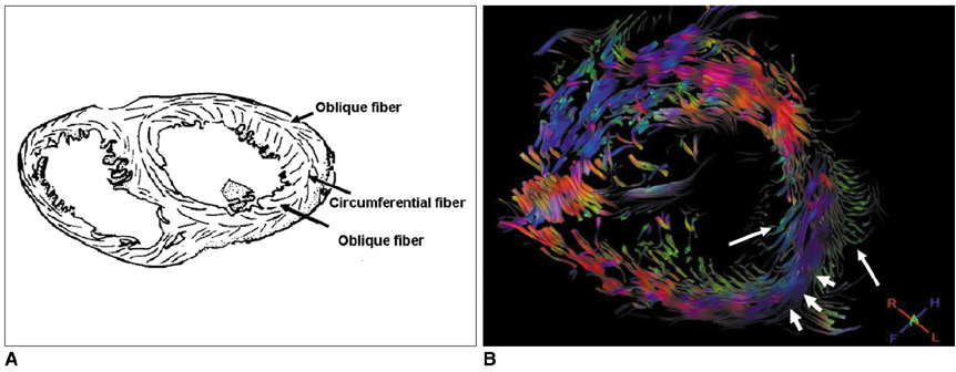
Fig. 3 Schematic cross section of heart at midventricular level (A, modified from ref. 3) and corresponding examples of fiber tractography using 32 diffusion gradient directions (B). Both oblique fibers (long arrows) and circumferential fibers (short arrow) are nicely demonstrated on diffusion-tensor image.
Reference
-
1. Rademakers FE, Rogers WJ, Guier WH, Hutchins GM, Siu CO, Weisfeldt ML, et al. Relation of regional cross-fiber shortening to wall thickening in the intact heart. Three-dimensional strain analysis by NMR tagging. Circulation. 1994. 89:1174–1182.2. Taber LA, Yang M, Podszus WW. Mechanics of ventricular torsion. J Biomech. 1996. 29:745–752.3. Spotnitz HM. Macrodesign, structure, and mechanics of the left ventricle. J Thorac Cardiovasc Surg. 2000. 119:1053–1057.4. Streeter DD Jr, Spotnitz HM, Patel DP, Ross J Jr, Sonnenblick EH. Fiber orientation in the canine left ventricle during diastole and systole. Circ Res. 1969. 24:339–347.5. Wu EX, Wu Y, Nicholls JM, Wang J, Liao S, Zhu S, et al. MR diffusion tensor imaging study of postinfarct myocardium structural remodeling in a porcine model. Magn Reson Med. 2007. 58:687–695.6. Edelman RR, Gaa J, Wedeen VJ, Loh E, Hare JM, Prasad P, et al. In vivo measurement of water diffusion in the human heart. Magn Reson Med. 1994. 32:423–428.7. Reese TG, Weisskoff RM, Smith RN, Rosen BR, Dinsmore RE, Wedeen VJ. Imaging myocardial fiber architecture in vivo with magnetic resonance. Magn Reson Med. 1995. 34:786–791.8. Tseng WY, Reese TG, Weisskoff RM, Wedeen VJ. Cardiac diffusion tensor MRI in vivo without strain correction. Magn Reson Med. 1999. 42:393–403.9. Holmes AA, Scollan DF, Winslow RL. Direct histological validation of diffusion tensor MRI in formaldehyde-fixed myocardium. Magn Reson Med. 2000. 44:157–161.10. Scollan DF, Holmes A, Winslow R, Forder J. Histological validation of myocardial microstructure obtained from diffusion tensor magnetic resonance imaging. Am J Physiol. 1998. 275:H2308–H2318.11. Tseng WY, Wedeen VJ, Reese TG, Smith RN, Halpern EF. Diffusion tensor MRI of myocardial fibers and sheets: correspondence with visible cut-face texture. J Magn Reson Imaging. 2003. 17:31–42.12. Wu MT, Tseng WY, Su MY, Liu CP, Chiou KR, Wedeen VJ, et al. Diffusion tensor magnetic resonance imaging mapping the fiber architecture remodeling in human myocardium after infarction: correlation with viability and wall motion. Circulation. 2006. 114:1036–1045.13. Lee JW, Kim JH, Kang HS, Lee JS, Choi JY, Yeom JS, et al. Optimization of acquisition parameters of diffusion-tensor magnetic resonance imaging in the spinal cord. Invest Radiol. 2006. 41:553–559.14. Wu EX, Wu Y, Tang H, Wang J, Yang J, Ng MC, et al. Study of myocardial fiber pathway using magnetic resonance diffusion tensor imaging. Magn Reson Imaging. 2007. 25:1048–1057.15. Tanenbaum LN. 3-T MR imaging: ready for clinical practice. AJNR Am J Neuroradiol. 2004. 25:1626–1627.16. Zhai G, Lin W, Wilber KP, Gerig G, Gilmore JH. Comparisons of regional white matter diffusion in healthy neonates and adults performed with a 3.0-T head-only MR imaging unit. Radiology. 2003. 229:673–681.17. Choi SI, Choi SH, Kim ST, Lim KH, Lim CH, Gong GY, et al. Irreversibly damaged myocardium at MR imaging with a necrotic tissue-specific contrast agent in a cat model. Radiology. 2000. 215:863–868.18. Jeong AK, Choi SI, Kim DH, Park SB, Lee SS, Choi SH, et al. Evaluation by contrast-enhanced MR imaging of the lateral border zone in reperfused myocardial infarction in a cat model. Korean J Radiol. 2001. 2:21–27.19. Jones DK. The effect of gradient sampling schemes on measures derived from diffusion tensor MRI: a Monte Carlo study. Magn Reson Med. 2004. 51:807–815.20. Okada T, Miki Y, Fushimi Y, Hanakawa T, Kanagaki M, Yamamoto A, et al. Diffusion-tensor fiber tractography: intraindividual comparison of 30-T and 15-T MR imaging. Radiology. 2006. 238:668–678.21. Jaermann T, Crelier G, Pruessmann KP, Golay X, Netsch T, van Muiswinkel AM, et al. SENSE-DTI at 3 T. Magn Reson Med. 2004. 51:230–236.22. Nagae-Poetscher LM, Jiang H, Wakana S, Golay X, van Zijl PC, Mori S. High-resolution diffusion tensor imaging of the brain stem at 3 T. AJNR Am J Neuroradiol. 2004. 25:1325–1330.23. Pruessmann KP, Weiger M, Scheidegger MB, Boesiger P. SENSE: sensitivity encoding for fast MRI. Magn Reson Med. 1999. 42:952–962.24. Cercignani M, Horsfield MA, Agosta F, Filippi M. Sensitivity-encoded diffusion tensor MR imaging of the cervical cord. AJNR Am J Neuroradiol. 2003. 24:1254–1256.25. Naganawa S, Koshikawa T, Kawai H, Fukatsu H, Ishigaki T, Maruyama K, et al. Optimization of diffusion-tensor MR imaging data acquisition parameters for brain fiber tracking using parallel imaging at 3 T. Eur Radiol. 2004. 14:234–238.26. Pierpaoli C, Basser PJ. Toward a quantitative assessment of diffusion anisotropy. Magn Reson Med. 1996. 36:893–906.27. Jiang Y, Guccione JM, Ratcliffe MB, Hsu EW. Transmural heterogeneity of diffusion anisotropy in the sheep myocardium characterized by MR diffusion tensor imaging. Am J Physiol Heart Circ Physiol. 2007. 293:H2377–H2384.28. Chang Y, Lee YJ, Kim YS, Kang DS, Sohn CH, Woo SK, et al. Histogram analysis of noise performance on fractional anisotropy brain MR image with different diffusion gradient numbers. J Korean Radiol Soc. 2005. 52:87–92. [Korean].
- Full Text Links
-
- Actions
-
Cited
- CITED
-
- Close
- Share
-
- Similar articles
-
- Anisotropy Measurement and Fiber Tracking of the White Matter by Using Diffusion Tensor MR Imaging: Influence of the Number of Diffusion-Sensitizing Gradient Direction
- Effect of Increasing Diffusion Gradient Direction Number on Diffusion Tensor Imaging Fiber Tracking in the Human Brain
- The Effect of Imaging Parameters of Diffusion Tensor Imaging on Fractional Anisotropy
- Histogram Analysis of Noise Performance on Fractional Anisotropy Brain MR Image with Different Diffusion Gradient Numbers
- Diffusion Tensor Imaging: Exploring the Motor Networks and Clinical Applications
