Repaired Supraspinatus Tendons in Clinically Improving Patients: Early Postoperative Findings and Interval Changes on MRI
- Affiliations
-
- 1Department of Radiology, Kyung Hee University Hospital, Seoul 130-702, Korea. francesca@daum.net
- 2Department of Orthopaedic Surgery, Kyung Hee University Hospital, Seoul 130-702, Korea.
- 3Department of Radiology, Kyung Hee University Hospital at Gangdong, Seoul 134-727, Korea.
- KMID: 2070181
- DOI: http://doi.org/10.3348/kjr.2015.16.2.363
Abstract
OBJECTIVE
To demonstrate and further determine the incidences of repaired supraspinatus tendons on early postoperative magnetic resonance imaging (MRI) findings in clinically improving patients and to evaluate interval changes on follow-up MRIs.
MATERIALS AND METHODS
Fifty patients, who showed symptomatic and functional improvements after supraspinatus tendon repair surgery and who underwent postoperative MRI twice with a time interval, were included. The first and the second postoperative MRIs were obtained a mean of 4.4 and 11.5 months after surgery, respectively. The signal intensity (SI) patterns of the repaired tendon on T2-weighted images from the first MRI were classified into three types of heterogeneous high SI with fluid-like bright high foci (type I), heterogeneous high SI without fluid-like bright high foci (type II), and heterogeneous or homogeneous low SI (type III). Interval changes in the SI pattern, tendon thickness, and rotator cuff interval thickness between the two postoperative MRIs were evaluated.
RESULTS
The SI patterns on the first MRI were type I or II in 45 tendons (90%) and type III in five (10%). SI decreased significantly on the second MRI (p < 0.050). The mean thickness of repaired tendons and rotator cuff intervals also decreased significantly (p < 0.050).
CONCLUSION
Repaired supraspinatus tendons exhibited high SI in 90% of clinically improving patients on MRI performed during the early postsurgical period. The increased SI and thickness of the repaired tendon decreased on the later MRI, suggesting a gradual healing process rather than a retear.
Keyword
MeSH Terms
Figure
-

Fig. 1 Signal intensity (SI) patterns of repaired tendons on oblique coronal T2-weighted images. A. Type I tendon (heterogeneously high SI with fluid-like bright high SI foci) (arrow). B. Type II tendon (heterogeneously high SI without fluid-like bright high SI foci). C. Type III tendon (heterogeneously or homogeneously low SI).
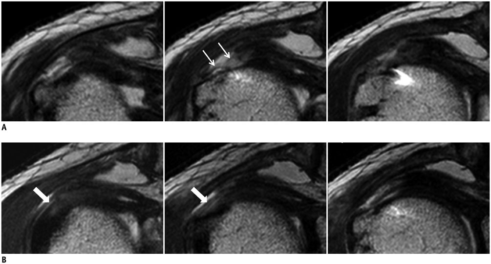
Fig. 2 65-year-old man with repaired tendon that changed from type I to type II during follow-up period. A. Supraspinatus tendon was type I pattern 4 months after surgery, including fluid-like bright high signal intensity (SI) foci on contiguous oblique coronal T2-weighted images. Linear or curvilinear structures with intermediate to low SI, which appeared to be suture material, are detected within high SI area at repair site (thin arrows). B. Previous fluid-like bright high SI focus is replaced by low SI striations (thick arrows) 8 months after surgery, and tendon shows heterogeneously high SI without fluid-like bright high SI, consistent with type II pattern.

Fig. 3 55-year-old woman with repaired tendon that changed from type I to type III during follow-up period. A. Supraspinatus tendon shows type I pattern with fluid-like bright high signal intensity (SI) foci (thick arrow) on oblique coronal T2-weighted images 5 months after surgery. Linear or curvilinear structures with intermediate to low SI, which appeared to be suture material, are detected within high SI area at repair site (thin arrows). B. Tendon shows type III pattern with homogeneously low SI 11 months after surgery.

Fig. 4 64-year-old man with repaired tendon that changed from type II to type III during follow-up period. A. Supraspinatus tendon shows type II pattern with heterogeneously high signal intensity (SI) (arrow) on oblique coronal T2-weighted image 4 months after surgery. B. Tendon shows type III pattern with decreases in both SI and thickness (arrow) 12 months after surgery.
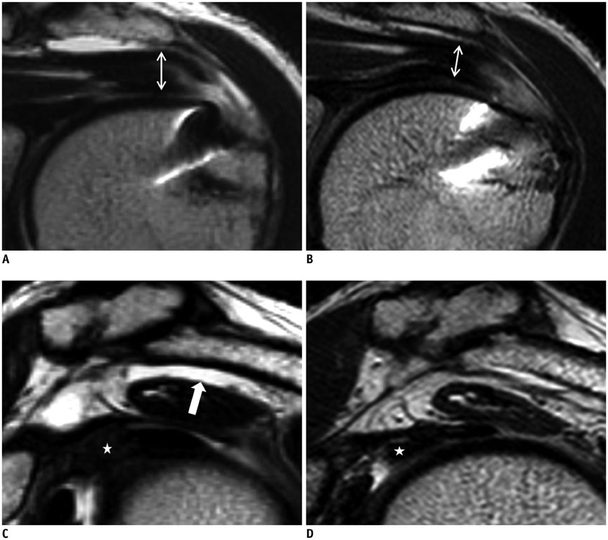
Fig. 5 52-year-old man with repaired tendon that changed from type I to type II during follow-up period, with decreased subacromial fluid and edema of rotator cuff interval. A, B. Serial oblique coronal T2-weighted images show thin supraspinatus tendon (double-headed arrow), and fluid SI of tendon disappears on second follow-up magnetic resonance image (MRI) (B) taken 6 months after first (A). C, D. Oblique sagittal images of first (C) and second (D) follow-up MRIs. Subacromial fluid (arrow in C) resolved, and thickness of rotator cuff interval (★ in C, D) decreased markedly.
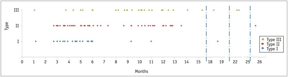
Fig. 6 Distribution of signal intensity (SI) patterns on 100 postoperative magnetic resonance image based on follow-up period.
Cited by 1 articles
-
Texture Analysis of Torn Rotator Cuff on Preoperative Magnetic Resonance Arthrography as a Predictor of Postoperative Tendon Status
Yeonah Kang, Guen Young Lee, Joon Woo Lee, Eugene Lee, Bohyoung Kim, Su Jin Kim, Joong Mo Ahn, Heung Sik Kang
Korean J Radiol. 2017;18(4):691-698. doi: 10.3348/kjr.2017.18.4.691.
Reference
-
1. Mohana-Borges AV, Chung CB, Resnick D. MR imaging and MR arthrography of the postoperative shoulder: spectrum of normal and abnormal findings. Radiographics. 2004; 24:69–85.2. Zlatkin MB. MRI of the postoperative shoulder. Skeletal Radiol. 2002; 31:63–80.3. Fealy S, Adler RS, Drakos MC, Kelly AM, Allen AA, Cordasco FA, et al. Patterns of vascular and anatomical response after rotator cuff repair. Am J Sports Med. 2006; 34:120–127.4. Crim J, Burks R, Manaster BJ, Hanrahan C, Hung M, Greis P. Temporal evolution of MRI findings after arthroscopic rotator cuff repair. AJR Am J Roentgenol. 2010; 195:1361–1366.5. Ellman H, Hanker G, Bayer M. Repair of the rotator cuff. End-result study of factors influencing reconstruction. J Bone Joint Surg Am. 1986; 68:1136–1114.6. Owen RS, Iannotti JP, Kneeland JB, Dalinka MK, Deren JA, Oleaga L. Shoulder after surgery: MR imaging with surgical validation. Radiology. 1993; 186:443–447.7. Thomazeau H, Boukobza E, Morcet N, Chaperon J, Langlais F. Prediction of rotator cuff repair results by magnetic resonance imaging. Clin Orthop Relat Res. 1997; (344):275–283.8. Oh JH, Kim SH, Ji HM, Jo KH, Bin SW, Gong HS. Prognostic factors affecting anatomic outcome of rotator cuff repair and correlation with functional outcome. Arthroscopy. 2009; 25:30–39.9. Gaenslen ES, Satterlee CC, Hinson GW. Magnetic resonance imaging for evaluation of failed repairs of the rotator cuff. Relationship to operative findings. J Bone Joint Surg Am. 1996; 78:1391–1139.10. Bryant L, Shnier R, Bryant C, Murrell GA. A comparison of clinical estimation, ultrasonography, magnetic resonance imaging, and arthroscopy in determining the size of rotator cuff tears. J Shoulder Elbow Surg. 2002; 11:219–224.11. Motamedi AR, Urrea LH, Hancock RE, Hawkins RJ, Ho C. Accuracy of magnetic resonance imaging in determining the presence and size of recurrent rotator cuff tears. J Shoulder Elbow Surg. 2002; 11:6–10.12. Schaefer O, Winterer J, Lohrmann C, Laubenberger J, Reichelt A, Langer M. Magnetic resonance imaging for supraspinatus muscle atrophy after cuff repair. Clin Orthop Relat Res. 2002; (403):93–99.13. Torstensen ET, Hollinshead RM. Comparison of magnetic resonance imaging and arthroscopy in the evaluation of shoulder pathology. J Shoulder Elbow Surg. 1999; 8:42–45.14. Magee TH, Gaenslen ES, Seitz R, Hinson GA, Wetzel LH. MR imaging of the shoulder after surgery. AJR Am J Roentgenol. 1997; 168:925–928.15. Boileau P, Brassart N, Watkinson DJ, Carles M, Hatzidakis AM, Krishnan SG. Arthroscopic repair of full-thickness tears of the supraspinatus: does the tendon really heal. J Bone Joint Surg Am. 2005; 87:1229–1240.16. Charousset C, Duranthon LD, Grimberg J, Bellaiche L. [Arthro-C-scan analysis of rotator cuff tears healing after arthroscopic repair: analysis of predictive factors in a consecutive series of 167 arthroscopic repairs]. Rev Chir Orthop Reparatrice Appar Mot. 2006; 92:223–233.17. Huijsmans PE, Pritchard MP, Berghs BM, van Rooyen KS, Wallace AL, de Beer JF. Arthroscopic rotator cuff repair with double-row fixation. J Bone Joint Surg Am. 2007; 89:1248–1257.18. Lafosse L, Brozska R, Toussaint B, Gobezie R. The outcome and structural integrity of arthroscopic rotator cuff repair with use of the double-row suture anchor technique. J Bone Joint Surg Am. 2007; 89:1533–1541.19. Sugaya H, Maeda K, Matsuki K, Moriishi J. Repair integrity and functional outcome after arthroscopic double-row rotator cuff repair. A prospective outcome study. J Bone Joint Surg Am. 2007; 89:953–996.20. Mellado JM, Calmet J, Olona M, Ballabriga J, Camins A, Pérez del, et al. MR assessment of the repaired rotator cuff: prevalence, size, location, and clinical relevance of tendon rerupture. Eur Radiol. 2006; 16:2186–2196.21. Klepps S, Bishop J, Lin J, Cahlon O, Strauss A, Hayes P, et al. Prospective evaluation of the effect of rotator cuff integrity on the outcome of open rotator cuff repairs. Am J Sports Med. 2004; 32:1716–1722.22. Spielmann AL, Forster BB, Kokan P, Hawkins RH, Janzen DL. Shoulder after rotator cuff repair: MR imaging findings in asymptomatic individuals--initial experience. Radiology. 1999; 213:705–708.23. Rand T, Freilinger W, Breitenseher M, Trattnig S, Garcia M, Landsiedl F, et al. Magnetic resonance arthrography (MRA) in the postoperative shoulder. Magn Reson Imaging. 1999; 17:843–850.24. Rand T, Trattnig S, Breitenseher M, Freilinger W, Cochole M, Imhof H. [MR arthrography of the shoulder joint in a postoperative patient sample]. Radiologe. 1996; 36:966–970.25. Fujikawa A, Kyoto Y, Kawaguchi M, Naoi Y, Ukegawa Y. Achilles tendon after percutaneous surgical repair: serial MRI observation of uncomplicated healing. AJR Am J Roentgenol. 2007; 189:1169–1174.26. Jost B, Zumstein M, Pfirrmann CW, Gerber C. Long-term outcome after structural failure of rotator cuff repairs. J Bone Joint Surg Am. 2006; 88:472–479.27. Rafii M, Firooznia H, Golimbu C, Weinreb J. Magnetic resonance imaging of glenohumeral instability. Magn Reson Imaging Clin N Am. 1993; 1:87–104.28. Farley TE, Neumann CH, Steinbach LS, Jahnke AJ, Petersen SS. Full-thickness tears of the rotator cuff of the shoulder: diagnosis with MR imaging. AJR Am J Roentgenol. 1992; 158:347–351.29. Needell SD, Zlatkin MB, Sher JS, Murphy BJ, Uribe JW. MR imaging of the rotator cuff: peritendinous and bone abnormalities in an asymptomatic population. AJR Am J Roentgenol. 1996; 166:863–867.30. Harryman DT 2nd, Mack LA, Wang KY, Jackins SE, Richardson ML, Matsen FA 3rd. Repairs of the rotator cuff. Correlation of functional results with integrity of the cuff. J Bone Joint Surg Am. 1991; 73:982–998.
- Full Text Links
-
- Actions
-
Cited
- CITED
-
- Close
- Share
-
- Similar articles
-
- Fatty Degeneration and Atrophy of Rotator Cuffs: Comparison of Immediate Postoperative MRI with Preoperative MRI
- MRI Follow-up Study After Arthroscopic Repair of Multiple Rotator Cuff Tendons
- Reliability of the Supraspinatus Muscle Thickness Measurement by Ultrasonography
- Intramuscular Lipoma of the Supraspinatus Muscle with Supraspinatus Tendon Partial Tear
- Postoperative Ultrasound Findings of the Rotator Cuff Tendon after Arthroscopic Repair of a Rotator Cuff Tear
