Intramuscular Lipoma of the Supraspinatus Muscle with Supraspinatus Tendon Partial Tear
- Affiliations
-
- 1Department of Orthopedic Surgery, Daegu Fatima Hospital, Daegu, Korea. nso1020@naver.com
- KMID: 2106738
- DOI: http://doi.org/10.4055/jkoa.2015.50.1.49
Abstract
- Infiltrating lipoma in supraspinatus muscle on the shoulder is very rare. We performed open excision and rotator cuff repair on a patient who had infiltrating lipoma in supraspinatus muscle with partial tear of the supraspinatus tendon. We achieved a satisfactory outcome on one-year follow-up magnetic resonance imaging. We report on the case with a review of the literature.
Keyword
MeSH Terms
Figure
-
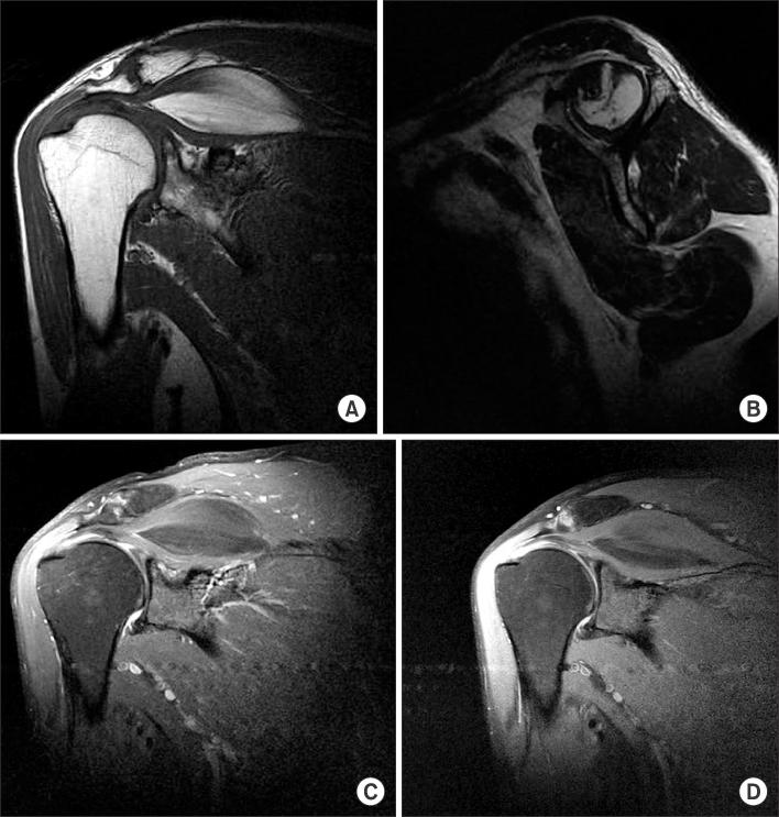
Figure 1 Coronal section on T1 shows the lipoma (A), sagittal section on T1 shows the lipoma (B), coronal section on T2 fat suppression image shows the lipoma (C). (D) Coronal section on T2 fat suppression image shows the supraspinatus muscle tendon parial tear.
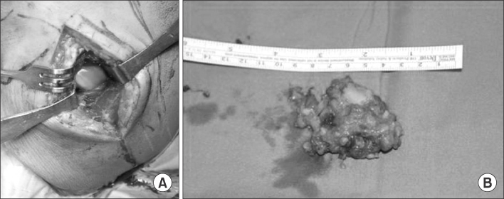
Figure 2 (A) Intraoperative photograph shows the lipoma. (B) Gross photograph shows the lipoma.
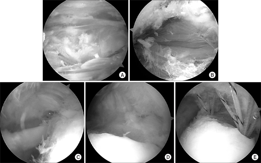
Figure 3 (A) Arthroscopic photography shows the lipoma. (B) Arthroscopic photography shows that the lipomas was removed from the supraspinatus muscle. (C) Arthroscopic photography shows the supraspinatus tendon tear. (D) Arthroscopic photography shows that a foot print of the supraspinatus. (E) A torn cuff was repaired by the transcuff method.
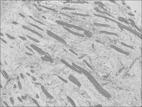
Figure 4 Histologic examination of a representative section of the mass shows a infiltrating lipoma characterized by mature adipocytes with muscle tissue (H&E, ×40).

Figure 5 (A) Follow-up magnetic resonance imaging (MRI) (postoperative one-year later) coronal section shows that remaining lipoma tissue (arrow). (B) Follow-up MRI (postoperative one-year later), sagittal section shows that remaining lipoma tissue. (C) Follow-up MRI (postoperative one-year later) coronal section shows that rotator cuff was repaired.
Reference
-
1. Neer CS 2nd. Impingement lesions. Clin Orthop Relat Res. 1983; 173:70–77.
Article2. Relwani J, Ogufere W, Orakwe S. Subacromial lipoma causing impingement syndrome of the shoulder: a case report. J Shoulder Elbow Surg. 2003; 12:202–203.
Article3. Egea Martínez JM, Mena JF. Lipoma of the supraspinatus muscle causing impingement syndrome: a case report. J Shoulder Elbow Surg. 2009; 18:e3–e5.
Article4. Ferrari L, Haynes P, Mack J, DiFelice GS. Intramuscular lipoma of the supraspinatus causing impingement syndrome. Orthopedics. 2009; 32:pii:orthosupersite.com/view.asp?rID=41927.
Article5. Weiss SW. Lipomatous tumors. Monogr Pathol. 1996; 38:207–239.6. Sungur N, Kilinç H, Ozdemr R, Sensöz O. An infiltrating intramuscular lipoma of the brachioradialis muscle. Ann Plast Surg. 2001; 46:353–354.
Article7. Oh JH, Gong HS, Kim HH. Paralysis of inferior branch of suprascapular nerve by a lipoma: a case report. J Korean Shoulder Elbow Soc. 2004; 7:103–107.
Article8. Matsumoto K, Hukuda S, Ishizawa M, Chano T, Okabe H. MRI findings in intramuscular lipomas. Skeletal Radiol. 1999; 28:145–152.
Article9. Blair B, Rokito AS, Cuomo F, Jarolem K, Zuckerman JD. Efficacy of injections of corticosteroids for subacromial impingement syndrome. J Bone Joint Surg Am. 1996; 78:1685–1689.
Article
- Full Text Links
-
- Actions
-
Cited
- CITED
-
- Close
- Share
-
- Similar articles
-
- Diagnosis of Partial Thickness Tear of Supraspinatus Tendon Using Dynamic Ultrasonography Under Resisted Scaption Position
- Partial-Thickness Tear of Supraspinatus and Infraspinatus Tendon Revisited: Based on MR Findings
- Isokinetic Muscle Strength and Muscle Endurance by the Types and Size of Rotator Cuff Tear in Men
- Sonoelastography on Supraspinatus Muscle-Tendon and Long Head of Biceps Tendon in Korean Professional Baseball Pitchers
- Plain Radiographic Findings of a Supraspinatus Tear: Correlation with MR Findings
