Magnetic Resonance Imaging-Guided Prostate Biopsy: Present and Future
- Affiliations
-
- 1Department of Radiology and Center for Imaging Science, Samsung Medical Center, Sungkyunkwan University School of Medicine, Seoul 135-710, Korea. chankyokim@skku.edu
- KMID: 2069987
- DOI: http://doi.org/10.3348/kjr.2015.16.1.90
Abstract
- Systemic transrectal ultrasound-guided biopsy (TRUSBx) is the standard procedure for diagnosing prostate cancer (PCa), but reveals a limited accuracy for the detection of cancer. Currently, multiparametric MR imaging (mp-MRI) is increasingly regarded as a promising method to detect PCa with an excellent positive predictive value. The use of mp-MRI during a MRI-guided biopsy (MRGB) procedure improves the quality of a targeted biopsy. The aim of this article is to provide an overview about the MRGB technique for PCa detection, to review the accuracy and clinical indications of MRGB and discuss its current issues and further directions. A MRGB seems accurate and efficient for the detection of clinically significant PCa in men with previous negative TRUSBx. Moreover, it may decrease the detection of clinically insignificant cancers with fewer biopsy cores.
Keyword
MeSH Terms
Figure
-

Fig. 1 56-year-old man with stage T3a Gleason 4 + 3 prostate cancer (prostate specific antigen = 28.5 ng/mL) in left peripheral zone (PZ). A. On axial T2-weighted image, focal mass of low signal intensity (arrow) is seen in left PZ on mid-gland level with indistinct and irregular capsular margin suggesting extracapsular extension. B, C. Mass (arrow) in left PZ shows focally high signal intensity on axial diffusion-weighted (B) and reduced apparent diffusion coefficient (ADC) value on ADC map (C) images. D. On axial color-coded ktrans map of dynamic contrast-enhanced image, mass (arrow) reveals focal, asymmetric enhancement in corresponding site of Figure 1A.
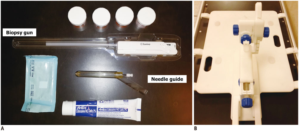
Fig. 2 In-bore targeting devices. A. 150/175 mm 18-gauge automatic biopsy gun, prostate needle guide, bottles for sampled cores and lidocaine jelly are shown. B. Biopsy device with base plate for patient positioning (Invivo, Schwerin, Germany).
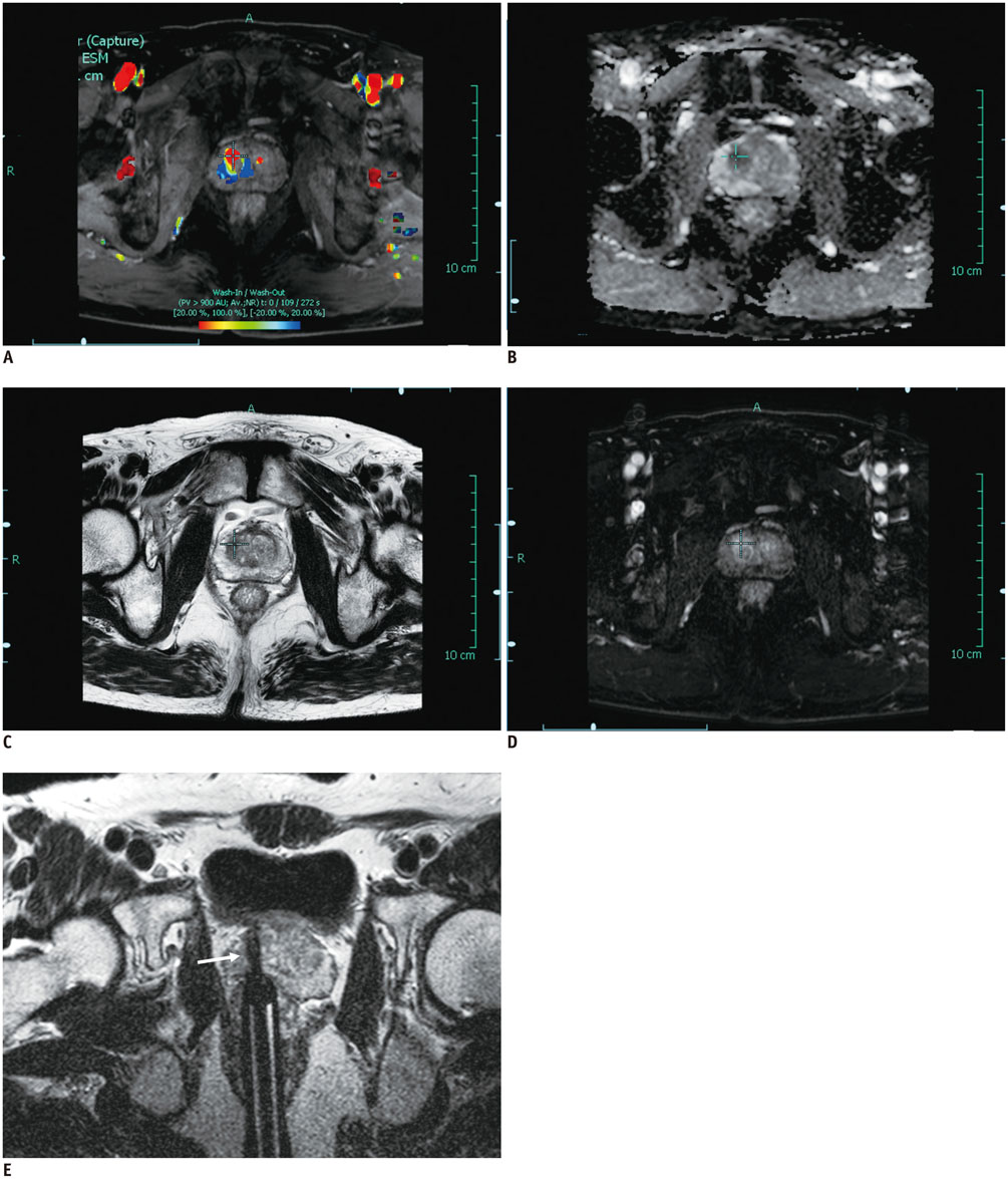
Fig. 3 74-year-old man with stage T2a Gleason score 3 + 4 prostate cancer (prostate specific antigen = 14.56 ng/mL) in right transition zone (TZ) with history of one previous negative transrectal ultrasound-guided biopsy underwent subsequent multiparametric MRI for clinical suspicion of prostate cancer. A-D. Axial color-coded wash-in/wash-out image (A) and dynamic contrast-enhanced (D) images show asymmetric increased enhancement (cross) in right TZ. This mass demonstrates focal low signal intensity (cross) on axial apparent diffusion coefficient map (B) and T2-weighted (C) images. E. Oblique axial T2-weighted image confirms needle position (arrow) in right TZ. In this MRI-guided biopsy specimen, Gleason score 3 + 4 prostate cancer was found.
Reference
-
1. Schröder FH, Roobol MJ. Defining the optimal prostatespecific antigen threshold for the diagnosis of prostate cancer. Curr Opin Urol. 2009; 19:227–231.2. Schröder FH, Carter HB, Wolters T, van den Bergh RC, Gosselaar C, Bangma CH, et al. Early detection of prostate cancer in 2007. Part 1: PSA and PSA kinetics. Eur Urol. 2008; 53:468–447.3. Schröder FH, van der Maas P, Beemsterboer P, Kruger AB, Hoedemaeker R, Rietbergen J, et al. Evaluation of the digital rectal examination as a screening test for prostate cancer. Rotterdam section of the European Randomized Study of Screening for Prostate Cancer. J Natl Cancer Inst. 1998; 90:1817–1182.4. Noguchi M, Stamey TA, McNeal JE, Yemoto CM. Relationship between systematic biopsies and histological features of 222 radical prostatectomy specimens: lack of prediction of tumor significance for men with nonpalpable prostate cancer. J Urol. 2001; 166:104–109. discussion 109-110.5. Hambrock T, Hoeks C, Hulsbergen-van de Kaa C, Scheenen T, Fütterer J, Bouwense S, et al. Prospective assessment of prostate cancer aggressiveness using 3-T diffusion-weighted magnetic resonance imaging-guided biopsies versus a systematic 10-core transrectal ultrasound prostate biopsy cohort. Eur Urol. 2012; 61:177–184.6. Hoeks CM, Barentsz JO, Hambrock T, Yakar D, Somford DM, Heijmink SW, et al. Prostate cancer: multiparametric MR imaging for detection, localization, and staging. Radiology. 2011; 261:46–66.7. Barentsz JO, Richenberg J, Clements R, Choyke P, Verma S, Villeirs G, et al. ESUR prostate MR guidelines 2012. Eur Radiol. 2012; 22:746–757.8. Kim CK, Park BK. Update of prostate magnetic resonance imaging at 3 T. J Comput Assist Tomogr. 2008; 32:163–172.9. Presti JC Jr, O'Dowd GJ, Miller MC, Mattu R, Veltri RW. Extended peripheral zone biopsy schemes increase cancer detection rates and minimize variance in prostate specific antigen and age related cancer rates: results of a community multi-practice study. J Urol. 2003; 169:125–129.10. Eskew LA, Bare RL, McCullough DL. Systematic 5 region prostate biopsy is superior to sextant method for diagnosing carcinoma of the prostate. J Urol. 1997; 157:199–202. discussion 202-203.11. Hricak H, Choyke PL, Eberhardt SC, Leibel SA, Scardino PT. Imaging prostate cancer: a multidisciplinary perspective. Radiology. 2007; 243:28–53.12. Ouzzane A, Puech P, Lemaitre L, Leroy X, Nevoux P, Betrouni N, et al. Combined multiparametric MRI and targeted biopsies improve anterior prostate cancer detection, staging, and grading. Urology. 2011; 78:1356–1362.13. Divrik RT, Eroglu A, Sahin A, Zorlu F, Ozen H. Increasing the number of biopsies increases the concordance of Gleason scores of needle biopsies and prostatectomy specimens. Urol Oncol. 2007; 25:376–382.14. Kvåle R, Møller B, Wahlqvist R, Fosså SD, Berner A, Busch C, et al. Concordance between Gleason scores of needle biopsies and radical prostatectomy specimens: a population-based study. BJU Int. 2009; 103:1647–1654.15. Wolters T, Roobol MJ, van Leeuwen PJ, van den Bergh RC, Hoedemaeker RF, van Leenders GJ, et al. A critical analysis of the tumor volume threshold for clinically insignificant prostate cancer using a data set of a randomized screening trial. J Urol. 2011; 185:121–125.16. Scattoni V, Zlotta A, Montironi R, Schulman C, Rigatti P, Montorsi F. Extended and saturation prostatic biopsy in the diagnosis and characterisation of prostate cancer: a critical analysis of the literature. Eur Urol. 2007; 52:1309–1322.17. Turkbey B, Mani H, Shah V, Rastinehad AR, Bernardo M, Pohida T, et al. Multiparametric 3T prostate magnetic resonance imaging to detect cancer: histopathological correlation using prostatectomy specimens processed in customized magnetic resonance imaging based molds. J Urol. 2011; 186:1818–1824.18. Rouse P, Shaw G, Ahmed HU, Freeman A, Allen C, Emberton M. Multi-parametric magnetic resonance imaging to rule-in and rule-out clinically important prostate cancer in men at risk: a cohort study. Urol Int. 2011; 87:49–53.19. Marks L, Young S, Natarajan S. MRI-ultrasound fusion for guidance of targeted prostate biopsy. Curr Opin Urol. 2013; 23:43–50.20. Park BK, Park JW, Park SY, Kim CK, Lee HM, Jeon SS, et al. Prospective evaluation of 3-T MRI performed before initial transrectal ultrasound-guided prostate biopsy in patients with high prostate-specific antigen and no previous biopsy. AJR Am J Roentgenol. 2011; 197:W876–W881.21. Puech P, Rouvière O, Renard-Penna R, Villers A, Devos P, Colombel M, et al. Prostate cancer diagnosis: multiparametric MR-targeted biopsy with cognitive and transrectal USMR fusion guidance versus systematic biopsy--prospective multicenter study. Radiology. 2013; 268:461–469.22. Kumar R, Nayyar R, Kumar V, Gupta NP, Hemal AK, Jagannathan NR, et al. Potential of magnetic resonance spectroscopic imaging in predicting absence of prostate cancer in men with serum prostate-specific antigen between 4 and 10 ng/ml: a follow-up study. Urology. 2008; 72:859–863.23. Rastinehad AR, Baccala AA Jr, Chung PH, Proano JM, Kruecker J, Xu S, et al. D'Amico risk stratification correlates with degree of suspicion of prostate cancer on multiparametric magnetic resonance imaging. J Urol. 2011; 185:815–820.24. Natarajan S, Marks LS, Margolis DJ, Huang J, Macairan ML, Lieu P, et al. Clinical application of a 3D ultrasound-guided prostate biopsy system. Urol Oncol. 2011; 29:334–342.25. Sonn GA, Natarajan S, Margolis DJ, MacAiran M, Lieu P, Huang J, et al. Targeted biopsy in the detection of prostate cancer using an office based magnetic resonance ultrasound fusion device. J Urol. 2013; 189:86–91.26. Pinto PA, Chung PH, Rastinehad AR, Baccala AA Jr, Kruecker J, Benjamin CJ, et al. Magnetic resonance imaging/ultrasound fusion guided prostate biopsy improves cancer detection following transrectal ultrasound biopsy and correlates with multiparametric magnetic resonance imaging. J Urol. 2011; 186:1281–1285.27. Hambrock T, Somford DM, Hoeks C, Bouwense SA, Huisman H, Yakar D, et al. Magnetic resonance imaging guided prostate biopsy in men with repeat negative biopsies and increased prostate specific antigen. J Urol. 2010; 183:520–527.28. Hoeks CM, Schouten MG, Bomers JG, Hoogendoorn SP, Hulsbergen-van de, Hambrock T, et al. Three-Tesla magnetic resonance-guided prostate biopsy in men with increased prostate-specific antigen and repeated, negative, random, systematic, transrectal ultrasound biopsies: detection of clinically significant prostate cancers. Eur Urol. 2012; 62:902–909.29. Pondman KM, Fütterer JJ, ten Haken B, Schultze Kool LJ, Witjes JA, Hambrock T, et al. MR-guided biopsy of the prostate: an overview of techniques and a systematic review. Eur Urol. 2008; 54:517–527.30. Franiel T, Stephan C, Erbersdobler A, Dietz E, Maxeiner A, Hell N, et al. Areas suspicious for prostate cancer: MR-guided biopsy in patients with at least one transrectal US-guided biopsy with a negative finding--multiparametric MR imaging for detection and biopsy planning. Radiology. 2011; 259:162–172.31. Somford DM, Hoeks CM, Hulsbergen-van de Kaa CA, Hambrock T, Fütterer JJ, Witjes JA, et al. Evaluation of diffusion-weighted MR imaging at inclusion in an active surveillance protocol for low-risk prostate cancer. Invest Radiol. 2013; 48:152–157.32. Vargas HA, Wassberg C, Akin O, Hricak H. MR imaging of treated prostate cancer. Radiology. 2012; 262:26–42.33. Rud E, Baco E, Lien D, Klotz D, Eggesbø HB. Detection of radiorecurrent prostate cancer using diffusion-weighted imaging and targeted biopsies. AJR Am J Roentgenol. 2014; 202:W241–W246.34. Moore CM, Robertson NL, Arsanious N, Middleton T, Villers A, Klotz L, et al. Image-guided prostate biopsy using magnetic resonance imaging-derived targets: a systematic review. Eur Urol. 2013; 63:125–140.35. Haffner J, Lemaitre L, Puech P, Haber GP, Leroy X, Jones JS, et al. Role of magnetic resonance imaging before initial biopsy: comparison of magnetic resonance imaging-targeted and systematic biopsy for significant prostate cancer detection. BJU Int. 2011; 108(8 Pt 2):E171–E178.36. Mian BM, Naya Y, Okihara K, Vakar-Lopez F, Troncoso P, Babaian RJ. Predictors of cancer in repeat extended multisite prostate biopsy in men with previous negative extended multisite biopsy. Urology. 2002; 60:836–840.37. Philip J, Hanchanale V, Foster CS, Javlé P. Importance of peripheral biopsies in maximising the detection of early prostate cancer in repeat 12-core biopsy protocols. BJU Int. 2006; 98:559–562.38. Roehl KA, Antenor JA, Catalona WJ. Serial biopsy results in prostate cancer screening study. J Urol. 2002; 167:2435–2439.39. Roethke M, Anastasiadis AG, Lichy M, Werner M, Wagner P, Kruck S, et al. MRI-guided prostate biopsy detects clinically significant cancer: analysis of a cohort of 100 patients after previous negative TRUS biopsy. World J Urol. 2012; 30:213–218.40. Sciarra A, Panebianco V, Ciccariello M, Salciccia S, Cattarino S, Lisi D, et al. Value of magnetic resonance spectroscopy imaging and dynamic contrast-enhanced imaging for detecting prostate cancer foci in men with prior negative biopsy. Clin Cancer Res. 2010; 16:1875–1883.41. Prando A, Kurhanewicz J, Borges AP, Oliveira EM Jr, Figueiredo E. Prostatic biopsy directed with endorectal MR spectroscopic imaging findings in patients with elevated prostate specific antigen levels and prior negative biopsy findings: early experience. Radiology. 2005; 236:903–910.42. Lee SH, Chung MS, Kim JH, Oh YT, Rha KH, Chung BH. Magnetic resonance imaging targeted biopsy in men with previously negative prostate biopsy results. J Endourol. 2012; 26:787–791.43. Brock M, Eggert T, Palisaar RJ, Roghmann F, Braun K, Löppenberg B, et al. Multiparametric ultrasound of the prostate: adding contrast enhanced ultrasound to real-time elastography to detect histopathologically confirmed cancer. J Urol. 2013; 189:93–98.44. Salomon G, Köllerman J, Thederan I, Chun FK, Budäus L, Schlomm T, et al. Evaluation of prostate cancer detection with ultrasound real-time elastography: a comparison with step section pathological analysis after radical prostatectomy. Eur Urol. 2008; 54:1354–1362.45. Woo S, Kim SY, Cho JY, Kim SH. Shear wave elastography for detection of prostate cancer: a preliminary study. Korean J Radiol. 2014; 15:346–355.46. Testa C, Schiavina R, Lodi R, Salizzoni E, Tonon C, D'Errico A, et al. Accuracy of MRI/MRSI-based transrectal ultrasound biopsy in peripheral and transition zones of the prostate gland in patients with prior negative biopsy. NMR Biomed. 2010; 23:1017–1026.
- Full Text Links
-
- Actions
-
Cited
- CITED
-
- Close
- Share
-
- Similar articles
-
- Medical imaging of prostate cancer
- Magnetic resonance imaging-transrectal ultrasound fusion image-guided prostate biopsy: Current status of the cancer detection and the prospects of tailor-made medicine of the prostate cancer
- MR-Guided Targeted Prostate Biopsy from Radiologists’ Perspective
- The Use of Magnetic Resonance Imaging in the Prostate Cancer Primary Diagnostic Pathway: Is It Ready for Primetime?
- Multiparametric MRI in the Detection of Clinically Significant Prostate Cancer
