Multiparametric MRI in the Detection of Clinically Significant Prostate Cancer
- Affiliations
-
- 1Department of Radiology and Nuclear Medicine, Radboud University Medical Center, Nijmegen 6500HB, the Netherlands. jurgen.futterer@Radboudumc.nl
- KMID: 2427228
- DOI: http://doi.org/10.3348/kjr.2017.18.4.597
Abstract
- Prostate cancer is the most common cancer among men aged 50 years and older in developed countries and the third leading cause of cancer-related death in men. Multiparametric prostate MR imaging is currently the most accurate imaging modality to detect, localize, and stage prostate cancer. The role of multi-parametric MR imaging in the detection of clinically significant prostate cancer are discussed. In addition, insights are provided in imaging techniques, protocol, and interpretation.
MeSH Terms
Figure
-
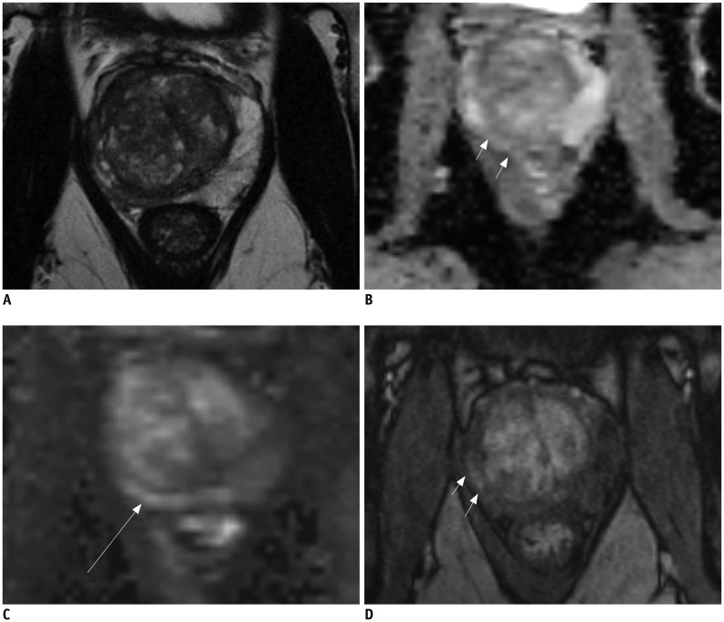
Fig. 1 61-year-old male with fluctuating PSA and current PSA level of 7.5 ng/mL.Prior biopsy session revealed no prostate cancer. A. T2-weighted MR imaging demonstrates asymmetric right peripheral zone with volume loss. Low signal intensity can be observed in right peripheral zone. B, C. ADC map (B) demonstrates intermediate signal intensity in right peripheral zone (arrows) and high b-value image (C) demonstrate intermediate to high signal intensity (arrow). D. T1-weighted post-contrast image demonstrates early uptake of contrast material in right peripheral zone (arrows). MR image guided biopsy revealed prostatitis and fibrosis. ADC = apparent diffusion coefficient, PSA = prostatespecific antigen
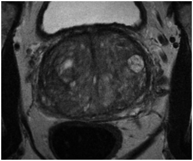
Fig. 2 75-year-old male with PSA level of 21 ng/mL.T2-weighted MR imaging reveals multiple BPH nodules (organized chaos) in transition zone. BPH = benign prostatic hyperplasia, PSA = prostate-specific antigen
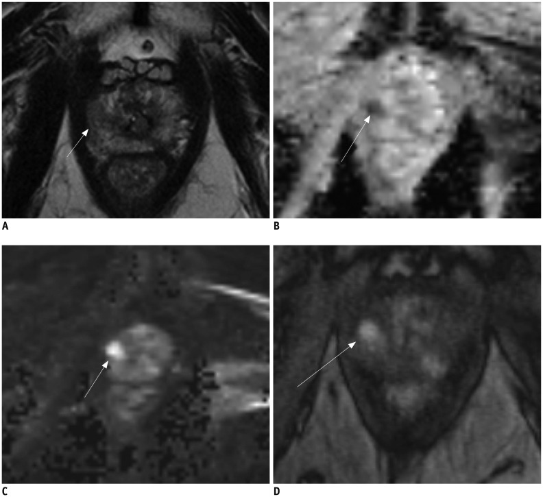
Fig. 3 62-year-old male with PSA level of 5.6 ng/mL.A. T2-weighted MR imaging reveals low signal intensity lesion (arrow) in right apex. B, C. ADC map (B) demonstrates distinct low signal intensity in right peripheral zone (arrow) and high b-value image (C) demonstrates high signal intensity (arrow). D. T1-weighted post-contrast image demonstrates early uptake of contrast material at corresponding location (arrow) (A-C). MR image targeted biopsy revealed Gleason 4 + 3 prostate cancer. ADC = apparent diffusion coefficient, PSA = prostate-specific antigen
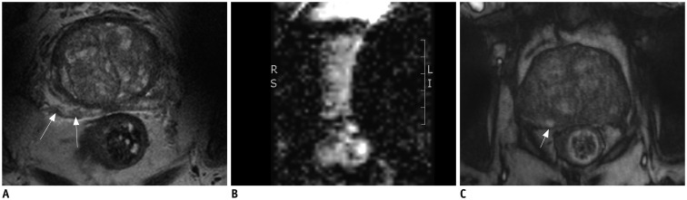
Fig. 4 78-year-old male with PSA level of 10.2 ng/mL and two negative prior transrectal ultrasound-guided biopsy sessions.A. T2-weighted MR imaging reveals low signal intensity lesion (arrows) in right peripheral zone. B. ADC map is not diagnostic as result of bilateral hip implants. C. T1-weighted post-contrast image demonstrates early uptake of contrast material at corresponding location (arrow) (A). MR image targeted biopsy revealed Gleason 3 + 4 prostate cancer. ADC = apparent diffusion coefficient, PSA = prostate-specific antigen
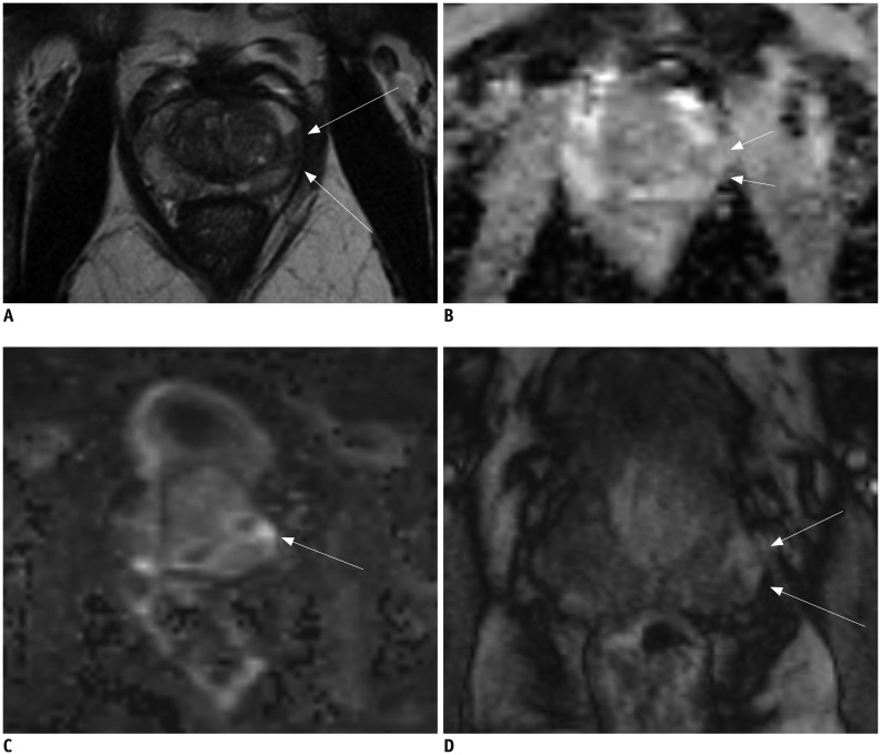
Fig. 5 68-year-old male with PSA level of 2.5 ng/mL.A. T2-weighted MR imaging reveals low signal intensity lesion (arrows) in left peripheral zone. B, C. ADC map (B) demonstrates distinct low signal intensity (arrows) and high b-value image (C) demonstrates high signal intensity at corresponding location (arrow) (A). D. T1-weighted post-contrast image demonstrates early uptake of contrast material (arrows) at corresponding location (A-C). MR image targeted biopsy revealed chronic prostatitis. ADC = apparent diffusion coefficient, PSA = prostate-specific antigen
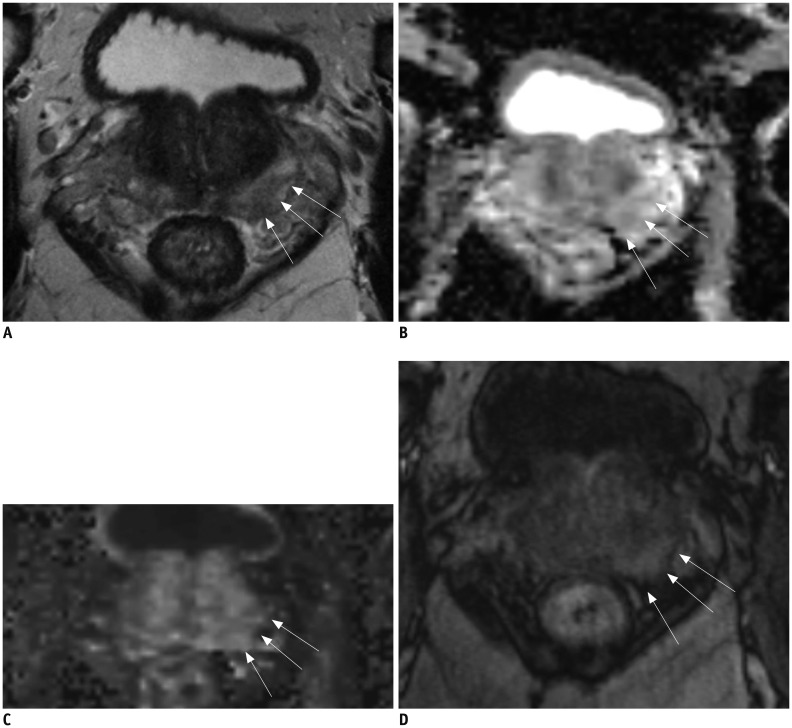
Fig. 6 64-year-old male with PSA level of 4.9 ng/mL.A. T2-weighted MR imaging reveals mild low signal intensity area (arrows) in left peripheral zone at base of prostate. B, C. ADC map (B) demonstrates mild low signal intensity (arrows) and high b-value image (C) demonstrates no high signal intensity at corresponding location (arrows) (A). D. T1-weighted post-contrast image demonstrates early uptake of contrast material (arrows) at corresponding location (A-C). Final PI-RADS assessment was 2. Clinical suspicion persisted and patient underwent MR image targeted biopsy which revealed Gleason 4 + 3 prostate cancer. ADC = apparent diffusion coefficient, PI-RADS = Prostate Imaging Reporting And Data System, PSA = prostate-specific antigen
Cited by 3 articles
-
Strategy for Prostate Cancer Patients with Low Prostate Specific Antigen Level (2.5 to 4.0 ng/mL)
Jae Hoon Chung, Jiwoong Yu, Wan Song, Minyong Kang, Hyun Hwan Sung, Hwang Gyun Jeon, Byong Chang Jeong, Seong IL Seo, Hyun Moo Lee, Seong Soo Jeon
J Korean Med Sci. 2020;35(41):e342. doi: 10.3346/jkms.2020.35.e342.Prostate Imaging-Reporting and Data System Version 2: Beyond Prostate Cancer Detection
Sung Yoon Park, Nam Hoon Cho, Dae Chul Jung, Young Taik Oh
Korean J Radiol. 2018;19(2):193-200. doi: 10.3348/kjr.2018.19.2.193.Yield of Repeat Targeted Direct in-Bore Magnetic Resonance-Guided Prostate Biopsy (MRGB) of the Same Lesions in Men Having a Prior Negative Targeted MRGB
Wulphert Venderink, Sjoerd FM Jenniskens, JP Michiel Sedelaar, Tsutomu Tamada, Jurgen J Fütterer
Korean J Radiol. 2018;19(4):733-741. doi: 10.3348/kjr.2018.19.4.733.
Reference
-
1. Ferlay J, Shin HR, Bray F, Forman D, Mathers C, Parkin DM. Estimates of worldwide burden of cancer in 2008: GLOBOCAN 2008. Int J Cancer. 2010; 127:2893–2917. PMID: 21351269.
Article2. Stamey TA, Caldwell M, McNeal JE, Nolley R, Hemenez M, Downs J. The prostate specific antigen era in the United States is over for prostate cancer: what happened in the last 20 years? J Urol. 2004; 172(4 Pt 1):1297–1301. PMID: 15371827.
Article3. Thompson IM, Pauler DK, Goodman PJ, Tangen CM, Lucia MS, Parnes HL, et al. Prevalence of prostate cancer among men with a prostate-specific antigen level < or =4.0 ng per milliliter. N Engl J Med. 2004; 350:2239–2246. PMID: 15163773.4. Djavan B, Ravery V, Zlotta A, Dobronski P, Dobrovits M, Fakhari M, et al. Prospective evaluation of prostate cancer detected on biopsies 1, 2, 3 and 4: when should we stop? J Urol. 2001; 166:1679–1683. PMID: 11586201.
Article5. Wang Y, Wang X, Yu J, Ouyang J, Shen W, Zhou Y, et al. Application of transrectal ultrasound-guided repeat needle biopsy in the diagnosis of prostate cancer in Chinese population: a retrospective study. J Res Med Sci. 2016; 21:79. PMID: 27904624.
Article6. Miyagawa T, Ishikawa S, Kimura T, Suetomi T, Tsutsumi M, Irie T, et al. Real-time virtual sonography for navigation during targeted prostate biopsy using magnetic resonance imaging data. Int J Urol. 2010; 17:855–860. PMID: 20807266.
Article7. Arsov C, Rabenalt R, Blondin D, Quentin M, Hiester A, Godehardt E, et al. Prospective randomized trial comparing magnetic resonance imaging (MRI)-guided in-bore biopsy to MRI-ultrasound fusion and transrectal ultrasound-guided prostate biopsy in patients with prior negative biopsies. Eur Urol. 2015; 68:713–720. PMID: 26116294.
Article8. Hoeks CM, Barentsz JO, Hambrock T, Yakar D, Somford DM, Heijmink SW, et al. Prostate cancer: multiparametric MR imaging for detection, localization, and staging. Radiology. 2011; 261:46–66. PMID: 21931141.
Article9. Barentsz JO, Richenberg J, Clements R, Choyke P, Verma S, Villeirs G, et al. ESUR prostate MR guidelines 2012. Eur Radiol. 2012; 22:746–757. PMID: 22322308.
Article10. Donati OF, Mazaheri Y, Afaq A, Vargas HA, Zheng J, Moskowitz CS, et al. Prostate cancer aggressiveness: assessment with whole-lesion histogram analysis of the apparent diffusion coefficient. Radiology. 2014; 271:143–152. PMID: 24475824.
Article11. Kobus T, Hambrock T, Hulsbergen-van de Kaa CA, Wright AJ, Barentsz JO, Heerschap A, et al. In vivo assessment of prostate cancer aggressiveness using magnetic resonance spectroscopic imaging at 3 T with an endorectal coil. Eur Urol. 2011; 60:1074–1080. PMID: 21419565.
Article12. Peng Y, Jiang Y, Yang C, Brown JB, Antic T, Sethi I, et al. Quantitative analysis of multiparametric prostate MR images: differentiation between prostate cancer and normal tissue and correlation with Gleason score--a computer-aided diagnosis development study. Radiology. 2013; 267:787–796. PMID: 23392430.
Article13. Weinreb JC, Barentsz JO, Choyke PL, Cornud F, Haider MA, Macura KJ, et al. PI-RADS prostate imaging-reporting and data system: 2015, version 2. Eur Urol. 2016; 69:16–40. PMID: 26427566.14. Vargas HA, Akin O, Franiel T, Goldman DA, Udo K, Touijer KA, et al. Normal central zone of the prostate and central zone involvement by prostate cancer: clinical and MR imaging implications. Radiology. 2012; 262:894–902. PMID: 22357889.
Article15. Ling D, Lee JK, Heiken JP, Balfe DM, Glazer HS, McClennan BL. Prostatic carcinoma and benign prostatic hyperplasia: inability of MR imaging to distinguish between the two diseases. Radiology. 1986; 158:103–107. PMID: 2416005.
Article16. Akin O, Sala E, Moskowitz CS, Kuroiwa K, Ishill NM, Pucar D, et al. Transition zone prostate cancers: features, detection, localization, and staging at endorectal MR imaging. Radiology. 2006; 239:784–792. PMID: 16569788.
Article17. desouza NM, Reinsberg SA, Scurr ED, Brewster JM, Payne GS. Magnetic resonance imaging in prostate cancer: the value of apparent diffusion coefficients for identifying malignant nodules. Br J Radiol. 2007; 80:90–95. PMID: 17303616.
Article18. Padhani AR, Harvey CJ, Cosgrove DO. Angiogenesis imaging in the management of prostate cancer. Nat Clin Pract Urol. 2005; 2:596–607. PMID: 16474547.
Article19. Collins DJ, Padhani AR. Dynamic magnetic resonance imaging of tumor perfusion. Approaches and biomedical challenges. IEEE Eng Med Biol Mag. 2004; 23:65–83.20. Epstein JI. Update on the Gleason grading system. Ann Pathol. 2011; 31(5 Suppl):S20–S26. PMID: 22054451.
Article21. Babaian RJ, Troncoso P, Bhadkamkar VA, Johnston DA. Analysis of clinicopathologic factors predicting outcome after radical prostatectomy. Cancer. 2001; 91:1414–1422. PMID: 11301387.
Article22. Zelefsky MJ, Kattan MW, Fearn P, Fearon BL, Stasi JP, Shippy AM, et al. Pretreatment nomogram predicting ten-year biochemical outcome of three-dimensional conformal radiotherapy and intensity-modulated radiotherapy for prostate cancer. Urology. 2007; 70:283–287. PMID: 17826490.
Article23. Fütterer JJ, Briganti A, De Visschere P, Emberton M, Giannarini G, Kirkham A, et al. Can clinically significant prostate cancer be detected with multiparametric magnetic resonance imaging? A systematic review of the literature. Eur Urol. 2015; 68:1045–1053. PMID: 25656808.
Article24. Chan TY, Partin AW, Walsh PC, Epstein JI. Prognostic significance of Gleason score 3+4 versus Gleason score 4+3 tumor at radical prostatectomy. Urology. 2000; 56:823–827. PMID: 11068310.
Article25. Vargas HA, Akin O, Franiel T, Mazaheri Y, Zheng J, Moskowitz C, et al. Diffusion-weighted endorectal MR imaging at 3 T for prostate cancer: tumor detection and assessment of aggressiveness. Radiology. 2011; 259:775–784. PMID: 21436085.
Article26. Rosenkrantz AB, Sigmund EE, Johnson G, Babb JS, Mussi TC, Melamed J, et al. Prostate cancer: feasibility and preliminary experience of a diffusional kurtosis model for detection and assessment of aggressiveness of peripheral zone cancer. Radiology. 2012; 264:126–135. PMID: 22550312.
Article27. Hambrock T, Somford DM, Huisman HJ, van Oort IM, Witjes JA, Hulsbergen-van de Kaa CA, et al. Relationship between apparent diffusion coefficients at 3.0-T MR imaging and Gleason grade in peripheral zone prostate cancer. Radiology. 2011; 259:453–461. PMID: 21502392.
Article28. Vos EK, Litjens GJ, Kobus T, Hambrock T, Hulsbergen-van de Kaa CA, Barentsz JO, et al. Assessment of prostate cancer aggressiveness using dynamic contrast-enhanced magnetic resonance imaging at 3 T. Eur Urol. 2013; 64:448–455. PMID: 23751135.
Article29. Oto A, Yang C, Kayhan A, Tretiakova M, Antic T, Schmid-Tannwald C, et al. Diffusion-weighted and dynamic contrast-enhanced MRI of prostate cancer: correlation of quantitative MR parameters with Gleason score and tumor angiogenesis. AJR Am J Roentgenol. 2011; 197:1382–1390. PMID: 22109293.
Article30. Bae H, Yoshida S, Matsuoka Y, Nakajima H, Ito E, Tanaka H, et al. Apparent diffusion coefficient value as a biomarker reflecting morphological and biological features of prostate cancer. Int Urol Nephrol. 2014; 46:555–561. PMID: 24022845.
Article31. Nagel KN, Schouten MG, Hambrock T, Litjens GJ, Hoeks CM, ten Haken B, et al. Differentiation of prostatitis and prostate cancer by using diffusion-weighted MR imaging and MR-guided biopsy at 3 T. Radiology. 2013; 267:164–172. PMID: 23329653.
Article32. Zakian KL, Sircar K, Hricak H, Chen HN, Shukla-Dave A, Eberhardt S, et al. Correlation of proton MR spectroscopic imaging with gleason score based on step-section pathologic analysis after radical prostatectomy. Radiology. 2005; 234:804–814. PMID: 15734935.
Article33. Wang L, Mazaheri Y, Zhang J, Ishill NM, Kuroiwa K, Hricak H. Assessment of biologic aggressiveness of prostate cancer: correlation of MR signal intensity with Gleason grade after radical prostatectomy. Radiology. 2008; 246:168–176. PMID: 18024440.
Article34. Lemaitre L, Puech P, Poncelet E, Bouyé S, Leroy X, Biserte J, et al. Dynamic contrast-enhanced MRI of anterior prostate cancer: morphometric assessment and correlation with radical prostatectomy findings. Eur Radiol. 2009; 19:470–480. PMID: 18758786.
Article35. Bratan F, Melodelima C, Souchon R, Hoang Dinh A, Mège-Lechevallier F, Crouzet S, et al. How accurate is multiparametric MR imaging in evaluation of prostate cancer volume? Radiology. 2015; 275:144–154. PMID: 25423145.
Article36. Lencioni R, Menchi I, Paolicchi A, Carini M, Amorosi A, Bartolozzi C. Prediction of pathological tumor volume in clinically localized prostate cancer: value of endorectal coil magnetic resonance imaging. MAGMA. 1997; 5:117–121. PMID: 9268075.
Article37. Coakley FV, Kurhanewicz J, Lu Y, Jones KD, Swanson MG, Chang SD, et al. Prostate cancer tumor volume: measurement with endorectal MR and MR spectroscopic imaging. Radiology. 2002; 223:91–97. PMID: 11930052.
Article38. Le Nobin J, Orczyk C, Deng FM, Melamed J, Rusinek H, Taneja SS, et al. Prostate tumour volumes: evaluation of the agreement between magnetic resonance imaging and histology using novel co-registration software. BJU Int. 2014; 114(6b):E105–E112. PMID: 24673731.
Article39. Anwar M, Westphalen AC, Jung AJ, Noworolski SM, Simko JP, Kurhanewicz J, et al. Role of endorectal MR imaging and MR spectroscopic imaging in defining treatable intraprostatic tumor foci in prostate cancer: quantitative analysis of imaging contour compared to whole-mount histopathology. Radiother Oncol. 2014; 110:303–308. PMID: 24444524.
Article40. Ouzzane A, Helfrich O, Le Nobin J, Puech P, Betrouni N, Villers A. Understanding the pathological implications of MRI: application to focal therapy planning. Curr Opin Urol. 2015; 25:198–204. PMID: 25768693.41. Fütterer JJ. MR imaging in local staging of prostate cancer. Eur J Radiol. 2007; 63:328–334. PMID: 17689908.
Article42. Fütterer JJ, Engelbrecht MR, Huisman HJ, Jager GJ, Hulsbergen-van De Kaa CA, Witjes JA, et al. Staging prostate cancer with dynamic contrast-enhanced endorectal MR imaging prior to radical prostatectomy: experienced versus less experienced readers. Radiology. 2005; 237:541–549. PMID: 16244263.
Article43. Vargas HA, Hötker AM, Goldman DA, Moskowitz CS, Gondo T, Matsumoto K, et al. Updated prostate imaging reporting and data system (PIRADS v2) recommendations for the detection of clinically significant prostate cancer using multiparametric MRI: critical evaluation using whole-mount pathology as standard of reference. Eur Radiol. 2016; 26:1606–1612. PMID: 26396111.
Article44. Lim CS, McInnes MD, Lim RS, Breau RH, Flood TA, Krishna S, et al. Prognostic value of Prostate Imaging and Data Reporting System (PI-RADS) v. 2 assessment categories 4 and 5 compared to histopathological outcomes after radical prostatectomy. Magn Reson Imaging. 2016; 11. 03. [Epub ahead of print]. DOI: 10.1002/jmri.25539.
Article45. Rosenkrantz AB, Ginocchio LA, Cornfeld D, Froemming AT, Gupta RT, Turkbey B, et al. Interobserver reproducibility of the PI-RADS version 2 lexicon: a multicenter study of six experienced prostate radiologists. Radiology. 2016; 280:793–804. PMID: 27035179.
Article46. Schoots IG, Roobol MJ, Nieboer D, Bangma CH, Steyerberg EW, Hunink MG. Magnetic resonance imaging-targeted biopsy may enhance the diagnostic accuracy of significant prostate cancer detection compared to standard transrectal ultrasound-guided biopsy: a systematic review and meta-analysis. Eur Urol. 2015; 68:438–450. PMID: 25480312.
Article47. de Rooij M, Hamoen EH, Fütterer JJ, Barentsz JO, Rovers MM. Accuracy of multiparametric MRI for prostate cancer detection: a meta-analysis. AJR Am J Roentgenol. 2014; 202:343–351. PMID: 24450675.
Article
- Full Text Links
-
- Actions
-
Cited
- CITED
-
- Close
- Share
-
- Similar articles
-
- Comparison of Multiparametric and Biparametric MRI in First Round Cognitive Targeted Prostate Biopsy in Patients with PSA Levels under 10 ng/mL
- Magnetic Resonance Imaging-Guided Prostate Biopsy: Present and Future
- MRI-Targeted Prostate Biopsy: What Radiologists Should Know
- Multiparametric MRI in Active Surveillance of Prostate Cancer: An Overview and a Practical Approach
- Multiparametric MRI of Prostate Cancer after Biopsy: Little Impact of Hemorrhage on Tumor Staging
