Early Gastric Cancer with Signet Ring Cell Histology Remained Unresected for 53 Months
- Affiliations
-
- 1Department of Surgery, Keimyung University School of Medicine, Daegu, Korea. sohnss@dsmc.or.kr
- KMID: 2055518
- DOI: http://doi.org/10.5230/jgc.2011.11.3.189
Abstract
- The natural course of untreated patients with signet ring cell carcinoma of the stomach remains poorly understood while assumptions have been made to distinguish it from other types of gastric cancer. A 74-year-old Korean woman was diagnosed with early gastric cancer with signet ring cell histology and refused surgery. A satellite lesion was identified 46 months after the initial diagnosis. The patient finally agreed to undergo distal subtotal gastrectomy 53 months following the initial diagnosis. Postoperative histological examination of both lesions confirmed signet ring cell carcinoma associated with submucosal invasion. There was no evidence of lymph node metastasis.
MeSH Terms
Figure
-
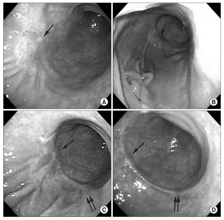
Fig. 1 Endoscopic images of the main lesion (single arrow) and the satellite lesion (double arrows) of early gastric cancer. The images were taken December 2005, when the initial diagnosis was made (A), subsequently on October 2006, 10 months after the diagnosis (B), on October 2009, 46 months after the diagnosis (C), and on March 2010, 51 months after the diagnosis (D).
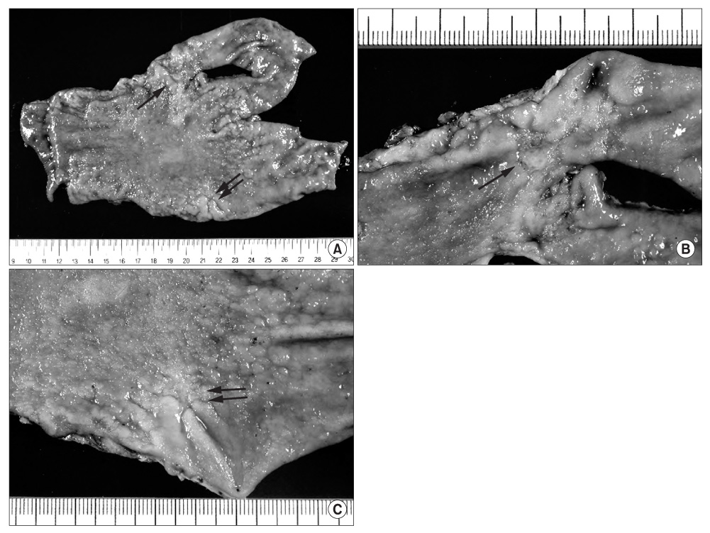
Fig. 2 The resected specimen in May 2010. The main lesion (single arrow) of type IIb early gastric cancer, 3.0×4.5 cm in size, was located in the anterior wall of the distal body of the stomach. The satellite lesion (double arrows) of a type IIb early gastric cancer, 2.0×2.5 cm in size, was located in the greater curvature of the distal body of the stomach. The two lesions were separated by normal mucosa. (A) Specimen. (B) Main lesion. (C) Satellite lesion.
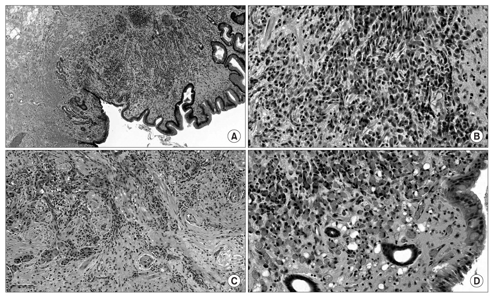
Fig. 3 Histopathologic findings. (A) The histopathologic findings of the main lesion were consistent with submucosal infiltration of tumor cells (H&E, ×200). (B) The tumor cells from the main lesion showed signet ring cell components (×400). (C) The histopathologic findings of the satellite lesion also showed submucosal infiltration of tumor cells (×200). (D) The tumor cells from the satellite lesion showed signet ring cell components as well (×400).
Reference
-
1. Maehara Y, Sakaguchi Y, Moriguchi S, Orita H, Korenaga D, Kohnoe S, et al. Signet ring cell carcinoma of the stomach. Cancer. 1992. 69:1645–1650.
Article2. Seo JH, Park JC, Kim YJ, Shin SK, Lee YC, Lee SK. Undifferentiated histology after endoscopic resection may predict synchronous and metachronous occurrence of early gastric cancer. Digestion. 2010. 81:35–42.
Article3. Reed VK, Krishnan S, Mansfield PF, Bhosale PR, Kim M, Das P, et al. Incidence, natural history, and patterns of locoregional recurrence in gastric cancer patients treated with preoperative chemoradiotherapy. Int J Radiat Oncol Biol Phys. 2008. 71:741–747.
Article4. Graham DY, Uemura N. Natural history of gastric cancer after Helicobacter pylori eradication in Japan: after endoscopic resection, after treatment of the general population, and naturally. Helicobacter. 2006. 11:139–143.
Article5. Tsukuma H, Mishima T, Oshima A. Prospective study of "early" gastric cancer. Int J Cancer. 1983. 31:421–426.
Article6. Tsukuma H, Oshima A, Narahara H, Morii T. Natural history of early gastric cancer: a non-concurrent, long term, follow up study. Gut. 2000. 47:618–621.
Article7. Guanrei Y, Songliang Q, He H, Guizen F. Natural history of early esophageal squamous carcinoma and early adenocarcinoma of the gastric cardia in the People's Republic of China. Endoscopy. 1988. 20:95–98.
Article8. Furukawa K, Yamada K, Konomi K, Tanaka M. Gastric carcinoma resected 95 months after being diagnosed: report of a case. Surg Today. 1994. 24:756–758.
Article9. Son HJ, Yoo MW, Kong SH, Ahn HS, Lee IK, Kim WH, et al. advanced gastric cancer that was curatively resected 78 months after being diagnosed: report of a case. J Korean Gastric Cancer Assoc. 2010. 10:40–44.
Article10. Yao K, Iwashita A, Tanabe H, Nagahama T, Matsui T, Ueki T, et al. Novel zoom endoscopy technique for diagnosis of small flat gastric cancer: a prospective, blind study. Clin Gastroenterol Hepatol. 2007. 5:869–878.
Article11. Yalamarthi S, Witherspoon P, McCole D, Auld CD. Missed diagnoses in patients with upper gastrointestinal cancers. Endoscopy. 2004. 36:874–879.
Article12. Voutilainen ME, Juhola MT. Evaluation of the diagnostic accuracy of gastroscopy to detect gastric tumours: clinicopathological features and prognosis of patients with gastric cancer missed on endoscopy. Eur J Gastroenterol Hepatol. 2005. 17:1345–1349.
Article13. Oohara T, Aono G, Ukawa S, Takezoe K, Johjima Y, Kurosaka H, et al. Clinical diagnosis of minute gastric cancer less than 5 mm in diameter. Cancer. 1984. 53:162–165.
Article14. Otsuji E, Kuriu Y, Ichikawa D, Okamoto K, Hagiwara A, Yamagishi H. Clinicopathologic characteristics and prognosis of synchronous multifocal gastric carcinomas. Am J Surg. 2005. 189:116–119.
Article15. Hyung WJ, Noh SH, Lee JH, Huh JJ, Lah KH, Choi SH, et al. Early gastric carcinoma with signet ring cell histology. Cancer. 2002. 94:78–83.
Article
- Full Text Links
-
- Actions
-
Cited
- CITED
-
- Close
- Share
-
- Similar articles
-
- CT Finding of Signet Ring Cell Carcinoma of the Stomach
- Liver Metastasis of Early Gastric Cancer with Mixed Histology after Endoscopic Submucosal Dissection
- Clinical Study of Signet Ring Cell Carcinomas of the Stomach
- Cytologic Heterogeneity of Signet Ring Cell Carcinoma of the Stomach: Histochemical and electron microscopic observations
- Two Cases of Histopathologically Advanced (Stage IV)Early Gastric Cancer
