Liver Metastasis of Early Gastric Cancer with Mixed Histology after Endoscopic Submucosal Dissection
- Affiliations
-
- 1Department of Internal Medicine, Gangneung Asan Hospital, University of Ulsan College of Medicine, Gangneung, Korea. sajahoooo@naver.com
- KMID: 2148574
- DOI: http://doi.org/10.5946/ce.2015.48.3.247
Abstract
- The Japanese Classification of Gastric Carcinoma histologically classifies endoscopically resected gastric cancer into differentiated and undifferentiated types according to the presence or absence of tubular structures on histology. The former includes papillary adenocarcinoma and tubular types, and the latter includes poorly differentiated adenocarcinoma, signet ring cell carcinoma and mucinous adenocarcinoma. However, gastric cancer sometimes contains a mixture of differentiated and undifferentiated components, and the clinical outcomes of the histological mixture are unknown, especially following endoscopic resection of early gastric cancer (EGC). This case was within the guideline indications for endoscopic submucosal resection (ESD), although it contained a partly signet ring cell carcinoma component; it recurred after 19 months with multiple lymph node and liver metastases. This case shows that additional surgical resection after ESD should be performed for patients with any mixed signet ring cell component, even in mild or moderately differentiated EGC.
MeSH Terms
Figure
-
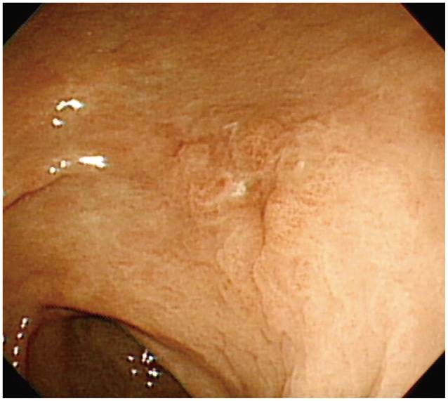
Fig. 1 Esophagogastroduodenoscopy showing IIa+IIc lesions on the posterior wall of the mid-antrum.

Fig. 2 En bloc esophagogastroduodenoscopy specimen; the carcinoma is confined within the red line.

Fig. 3 Pathologic findings. (A) Moderately differentiated area with signet ring cell features invades into the muscularis mucosa (H&E stain, ×100). (B) A clear vertical resection margin (H&E stain, ×100).
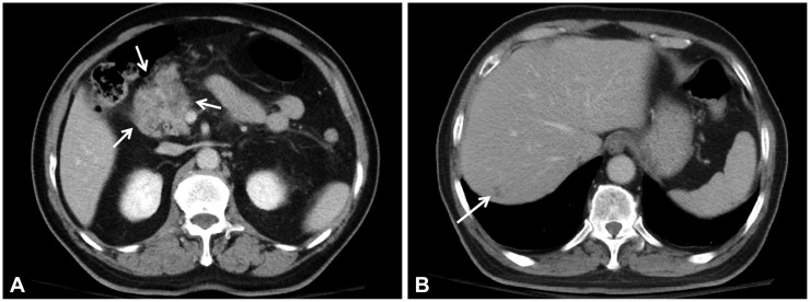
Fig. 4 Abdominal computed tomography findings. (A) A 3×5-cm necrotic mass (arrows) between the prepyloric antrum and pancreas head. (B) A 1.5-cm ill-defined mass (arrow) in segment 7 of the liver.
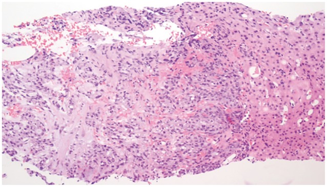
Fig. 5 Liver biopsy. A moderately differentiated adenocarcinoma with signet ring cell features compatible with liver metastasis of early gastric cancer (H&E stain, ×100).
Reference
-
1. Gotoda T, Jung HY. Endoscopic resection (endoscopic mucosal resection/ endoscopic submucosal dissection) for early gastric cancer. Dig Endosc. 2013; 25(Suppl 1):55–63. PMID: 23362925.
Article2. Coda S, Lee SY, Gotoda T. Endoscopic mucosal resection and endoscopic submucosal dissection as treatments for early gastrointestinal cancers in Western countries. Gut Liver. 2007; 1:12–21. PMID: 20485653.
Article3. Gotoda T. Endoscopic resection of early gastric cancer. Gastric Cancer. 2007; 10:1–11. PMID: 17334711.
Article4. Gotoda T, Yanagisawa A, Sasako M, et al. Incidence of lymph node metastasis from early gastric cancer: estimation with a large number of cases at two large centers. Gastric Cancer. 2000; 3:219–225. PMID: 11984739.
Article5. Japanese Gastric Cancer Association. Japanese Classification of Gastric Carcinoma: 2nd English edition. Gastric Cancer. 1998; 1:10–24. PMID: 11957040.6. Iwamoto J, Mizokami Y, Ito M, et al. Clinicopathological features of undifferentiated mixed type early gastric cancer treated with endoscopic submucosal dissection. Hepatogastroenterology. 2010; 57:185–190. PMID: 20422899.7. Shimizu H, Ichikawa D, Komatsu S, et al. The decision criterion of histological mixed type in "T1/T2" gastric carcinoma: comparison between TNM classification and Japanese Classification of Gastric Cancer. J Surg Oncol. 2012; 105:800–804. PMID: 22189799.8. Choi MH, Hong SJ, Han JP, et al. Therapeutic outcomes of endoscopic submucosal dissection in undifferentiated-type early gastric cancer. Korean J Gastroenterol. 2013; 61:196–202. PMID: 23624733.
Article9. Terada T. Primary signet-ring cell carcinoma of the pancreas diagnosed by endoscopic retrograde pancreatic duct biopsy: a case report with an immunohistochemical study. Endoscopy. 2012; 44(Suppl 2 UCTN):E141–E142. PMID: 22619039.
Article10. Japanese Gastric Cancer Association. Japanese gastric cancer treatment guidelines 2010 (ver. 3). Gastric Cancer. 2011; 14:113–123. PMID: 21573742.
- Full Text Links
-
- Actions
-
Cited
- CITED
-
- Close
- Share
-
- Similar articles
-
- Endoscopic Resection of Early Gastric Cancer
- Endoscopic Submucosal Dissection of Early Gastric Cancer
- Endoscopic Treatment for Early Gastric Cancer
- Future Development of Endoscopic Accessories for Endoscopic Submucosal Dissection
- Pitfalls in the Interpretation of Publications about Endoscopic Submucosal Dissection of Early Gastric Cancer with Undifferentiated-Type Histology
