Traumatic and Non-traumatic Osteonecrosis in the Femoral Head of a Rabbit Model
- Affiliations
-
- 1Laboratory of Veterinary Surgery, College of Veterinary Medicine, Chungbuk National University, Cheongju, Republic of Korea. ghkim@cbu.ac.kr
- KMID: 2053661
- DOI: http://doi.org/10.5625/lar.2011.27.2.127
Abstract
- Osteonecrosis of the femoral head is an idiopathic, debilitating and progressive disease. A number of traumatic or non-traumatic animal models have been reported for research on osteonecrosis. This study was performed to compare the efficacy of femoral head osteonecrosis in rabbits by traumatic and non-traumatic methods. Twenty-seven New Zealand White rabbits were divided into three experimental groups, nine heads each. Two groups were surgically induced into osteonecrosis; a steel cerclage wire was ligated tightly around the neck of the right femoral head (Group W), and the femoral neck was tied with a cerclage wire in the same way as in the W group, and burned by attachment of an electrode tip to the wire and then the wire was removed (Group B). The other group was induced into osteonecrosis with a single intra-muscular injection of 20 mg/kg methyl-prednisolone acetate single injection (Group M). In the control group, the left femoral head of animals in group W and B was used. After two weeks, rabbits were sacrificed and the femoral head and neck were collected. Osteonecrosis of the femoral head was evaluated by radiography, histology and immunohistology methods. Osteonecrosis lesions in the femoral head were identified in traumatic models of groups W and B. Cartilage degeneration in the superficial layer and TUNEL positive cells in the femoral head were detected more in Group B than in Group W. These findings revealed that short-term induced osteonecrosis of the femoral head was effectively achieved by cautery around the femoral neck.
Keyword
MeSH Terms
Figure
-
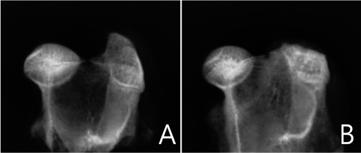
Figure 1 Digital radiograghic appearances of the normal (A) and osteonecrotic (B) femoral head. Radiodensity is increased in the osteonecrotic femoral head. No collapse or change was observed in femoral head sphericity.
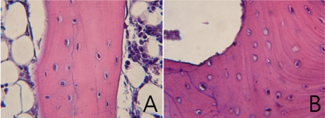
Figure 2 Histology of the epiphyseal region in the femoral head (H&E stain). Magnification ×400. A. Normal femoral head in the control group shows well stained nuclei of osteocytes and hematopoietic tissue, and normal trabecular bone and bone marrow. B. Osteonecrotic lesions in the femoral head of group B. The osteonecrotic lucanae are empty. Necrotic cells were observed in intertrabecular spaces.
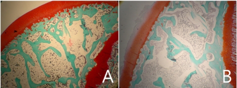
Figure 3 Histology of the epiphyseal cartilage region in the femoral head (Safranin O stain). Magnification ×400. A. Articular cartilage of the normal femoral head. B. Degenerated articular cartilage with fibrillation and loss of glycosaminoglycans of the femoral head in group B.
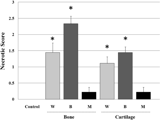
Figure 4 The average necrotic score of each group in trabecular bone and cartilage of the femoral heads. W group: wiring induced group, B group: burning induced group, M group: steroid induced group. Necrotic bone and cartilage changes were found to be significantly high in the W group and B group, compared with the control and M group (P<0.05).
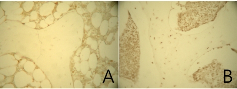
Figure 5 TUNEL labeling of the epiphyseal trabecular bone in the femoral head. Magnification ×400. A. No TUNEL positive cells or bone marrow cells were observed in the normal femoral head of the control group. B. TUNEL positive cells were observed. The TUNEL reaction shows apoptotic bodies in osteocytes and bone marrow cells.
Reference
-
1. Mont MA, Hungerford DS. Non-traumatic avascular necrosis of the femoral head. J Bone Joint Surg Am. 1995; 77:459–474. PMID: 7890797.
Article2. Trueta J. The normal vascular anatomy of the human femoral head during growth. J Bone Joint Surg Br. 1957; 39-B:358–394. PMID: 13438980.
Article3. Rokkanen P, Slatis P. Effect of compression on the healing of subcapital osteotomies of the femoral neck and on the avascularized femoral head. An experimental study on adult rabbits. Acta Orthop Scand. 1967; 38:163–173. PMID: 6033411.4. Kenzora JE, Steele RE, Yosipovitch ZH. Experimental osteonecrosis of the femoral head in adult rabbits. Clin Orthop. 1978; 130:8–46. PMID: 639409.
Article5. Rösingh GE, James J. Early phases of avascular necrosis of the femoral head in rabbits. J Bone Joint Surg Br. 1969; 51:165–174. PMID: 4179709.6. Robichon J, Desjardins JP, Koch M, Hooper CE. The femoral neck in Legg-Perthes' disease. Its relationship to epiphysial change and its importance in early prognosis. J Bone Joint Surg Br. 1974; 56:62–68. PMID: 4818856.7. Hofstaetter JG, Wang J, Yan J, Glimcher MJ. Changes in bone microarchitecture and bone mineral density following experimental oteonecrosis of the hip in rabbits. Cells Tissues Organs. 2006; 184:138–147. PMID: 17409739.8. Yamako G, Tokunaga K, Takano R, Endo N, Hara T. Morphological and mechanical evaluation of the cancellous bone in the rat femoral head after traumatic osteonecrosis. J Biomech Sci Eng. 2006; 1:195–203.
Article9. Kim J, Park J, Choi SH, Kim G. A comparison of surgical methods of inducing femoral head osteonecrosis in rats. J Vet Clin. 2010; 27:240–245.10. Norman D, Reis D, Zinman C, Misselevich I, Boss JH. Vascular deprivation-induced necrosis of the femoral head of the rat. An experimental model of avascular osteonecrosis in the skeletally immature individual or Legg-Perthes disease. Int J Exp Pathol. 1998; 79:173–181. PMID: 9741359.
Article11. Yamamoto T, Irisa T, Sugioka Y, Sueishi K. Effect of pulse methylprednisolone on bone and marrow tissue: corticosteroid-induced osteonecrosis in rabbits. Arthritis Rheum. 1997; 40:2055–2064. PMID: 9365096.12. Yang S, Wu X, Mei R, Yang C, Li J, Xu W, Ye S. Biomaterial-loaded allograft threaded cage for the treatment of femoral head osteonecrosis in a goat model. Biotechnol Bioeng. 2008; 100:560–566. PMID: 18438884.
Article13. Hernigou P, Poignard A, Nogier A, Manicom O. Fate of very small asymptomatic stage-osteonecrotic lesions of the hip. J Bone Joint Surg. 2004; 86:2589–2593. PMID: 15590840.14. Komiyama T, Nishida K, Yorimitsu M, Doi H, Miyazawa S, Kitamura A, Yoshida A, Nasu Y, Abe N, Ozaki T. Decreased levels of insulin-like growth Factor-1 and vascular endothelial growth factor relevant to the ossification disturbance in femoral heads spontaneous hypertensive rats. Acta Med Okayama. 2006; 60:141–148. PMID: 16838042.15. Levin D, Norman D, Zinman C, Rubinstein L, Sabo E, Misselevich I, Reis D, Boss JH. Treatment of experimental avascular necrosis of the femoral head with hyperbaric oxygen in rats. Exp Mol Pathol. 1999; 67:99–108. PMID: 10527761.16. Takaoka K, Yoshioka T, Hosoya T, Ono K, Takase T. The repair process in experimentally induced avascular necrosis of the femoral heads in dogs. Arch Orthop Trauma Surg. 1981; 99:109–115. PMID: 7316705.17. Mankin HJ. Nontraumatic necrosis of bone (osteonecrosis). N Engl J Med. 1992; 326:1473–1479. PMID: 1574093.
Article18. Arlet J. Nontraumatic avascular necrosis of the femoral head: past, present and future. Clin Orthop. 1992; 277:12–21. PMID: 1555331.
- Full Text Links
-
- Actions
-
Cited
- CITED
-
- Close
- Share
-
- Similar articles
-
- Osteonecrosis of Femoral Head after Pelvic Fracture: A Case Report
- Osteonecrosis of the Femoral Head in the Setting of a Complex Acetabulum Fracture without Hip Dislocation Treated Surgically Using Ilio-inguinal Approach: A Case Report
- Total Hip Arthroplasty in Patients with Avascular Necrosis of the Entire Femur
- Apoptosis in the Osteonecrosis of the Femoral Head
- The Early diagnostic Significance of Bone Marrow Pressure in Osteonecrosis of the Femoral Head
