Clin Endosc.
2012 Mar;45(1):99-102. 10.5946/ce.2012.45.1.99.
An Impacted Pancreatic Stone in the Papilla Induced Acute Obstructive Cholangitis in a Patient with Chronic Pancreatitis
- Affiliations
-
- 1Digestive Disease Center, CHA Bundang Medical Center, CHA University, Seongnam, Korea. endoscopy@cha.ac.kr
- KMID: 2048840
- DOI: http://doi.org/10.5946/ce.2012.45.1.99
Abstract
- Obstructive jaundice is very rarely caused by impaction of a pancreatic stone in the papilla. We report here on a case of obstructive jaundice with acute cholangitis that was caused by an impacted pancreatic stone in the papilla in a patient with chronic pancreatitis. A 48-year-old man presented with acute obstructive cholangitis. Abdominal computed tomography with the reconstructed image revealed distal biliary obstruction that was caused by a pancreatic stone in the pancreatic head, and there was also pancreatic ductal dilatation and parenchymal atrophy of the pancreatic body and tail with multiple calcifications. Emergency duodenoscopy revealed an impacted pancreatic stone in the papilla. Precut papillotomy using a needle knife was performed, followed by removal of the pancreatic stone using grasping forceps. After additional sphincterotomy, a large amount of dark-greenish bile juice gushed out. The patient rapidly improved and he has remained well.
Keyword
MeSH Terms
Figure
-

Fig. 1 Abdominal computed tomographic scanning with the reconstructed image revealed distal biliary obstruction by pancreatic stone in the pancreatic head, pancreatic ductal dilatation and parenchymal atrophy of the pancreatic body and tail with multiple calcifications. The diameter of the impacted stone in the pancreatic head was measured to be about 1 cm.
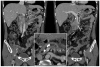
Fig. 2 The computed tomographic scanning with the reconstructive image performed 3 years previously.

Fig. 3 The duodenoscopic images. A whitish-yellow stone was exposed at the site of excision by performing precut papillotomy using a needle knife and the stone was removed by grasping forceps.
Reference
-
1. Moritomo H, Nakaya S, Takeyama Y, et al. A case of obstructive jaundice caused by incarceration of pancreatic stones in the ampulla of papilla Vater. Nihon Geka Gakkai Zasshi. 1990; 91:146–149. PMID: 2314379.2. Naik A, Shah SH, Relekar RG, Bapat RD. Pancreatic calculus causing obstructive jaundice. Indian J Gastroenterol. 1991; 10:27–28. PMID: 2004800.3. Little TE, Kozarek RA. Pancreatic stones as a cause of bile duct and ampullary obstruction: endoscopic treatment approaches. Gastrointest Endosc. 1993; 39:709–712. PMID: 8224700.
Article4. Hernandez JA, Zuckerman MJ, Moldes O. Pancreatic stone presenting with biliary obstruction. Gastrointest Endosc. 1994; 40:521–523. PMID: 7926556.
Article5. Kinoshita H, Imayama H, Sou H, et al. A case of obstructive icterus caused by incarceration of a pancreatic stone in the common channel of the pancreatobiliary ducts. Kurume Med J. 1996; 43:79–85. PMID: 8709563.
Article6. Naitoh I, Nakazawa T, Ohara H, et al. A case of obstructive jaundice caused by impaction of a pancreatic stone in the papilla for which a needle knife precut papillotomy was effective. JOP. 2008; 9:520–525. PMID: 18648146.7. Delhaye M, Arvanitakis M, Bali M, Matos C, Devière J. Endoscopic therapy for chronic pancreatitis. Scand J Surg. 2005; 94:143–153. PMID: 16111097.
Article8. Scott J, Summerfield JA, Elias E, Dick R, Sherlock S. Chronic pancreatitis: a cause of cholestasis. Gut. 1977; 18:196–201. PMID: 856677.
Article9. Sarles H, Sahel J. Cholestasis and lesions of the biliary tract in chronic pancreatitis. Gut. 1978; 19:851–857. PMID: 361513.
Article10. Sarles H, Sarles JC, Camatte R, et al. Observations on 205 confirmed cases of acute pancreatitis, recurring pancreatitis, and chronic pancreatitis. Gut. 1965; 6:545–559. PMID: 5857891.
Article11. Devière J, Devaere S, Baize M, Cremer M. Endoscopic biliary drainage in chronic pancreatitis. Gastrointest Endosc. 1990; 36:96–100. PMID: 2335299.
Article
- Full Text Links
-
- Actions
-
Cited
- CITED
-
- Close
- Share
-
- Similar articles
-
- An Impacted Pancreatic Stone at the Orifice of the Minor Papilla Causing a Bout of Acute Pancreatitis in a Patient with Pancreas Divisum
- Acute cholangitis and pancreatitis due to impacted papillary stone
- Case Review of Impacted Bile Duct Stone at Duodenal Papilla: Detection and Endoscopic Treatment
- A Case of Autoimmune Pancreatitis Manifested by a Pseudocyst and IgG4-Associated Cholangitis
- Acute Pancreatitis Induced by Compression of Main Pancreatic Duct due to Large Stones and Catheter in the Common Bile Duct
