Primary Spinal Cord Melanoma in Thoracic Spine with Leptomeningeal Dissemination and Presenting Hydrocephalus
- Affiliations
-
- 1Department of Neurosurgery, Ajou University Hospital, Ajou University School of Medicine, Suwon, Korea. nkyou@ajou.ac.kr
- KMID: 2048487
- DOI: http://doi.org/10.14791/btrt.2013.1.2.116
Abstract
- Primary spinal cord melanoma is a rare central nervous system malignant tumor. Usually it resembles an intradural extramedullary (IDEM) nerve sheath tumor or melanoma. We experienced a patient with upper thoracic primary IDEM spinal cord melanoma who was diagnosed to be with hydrocephalus and without intracranial lesions. Initial symptoms of the patient were related to the hydrocephalus and the primary spinal cord melanoma was diagnosed eight months later. At the first operation, complete resection was impossible and the patient refused additional radiotherapy or chemotherapy. At 22 months after surgery, the patient revisited our institution with recurrent both leg weakness. Leptomeningeal dissemination was present in the whole spinal cord and only partial resection of tumor was performed. The symptoms slightly improved after surgery. Primary spinal cord melanoma is extremely rare but complete resection and additional radiotherapy or chemotherapy can prolong the disease free interval. Hydrocephalus or signs of increased intracranial pressure may be the diagnostic clue of spinal cord malignancy and progression.
MeSH Terms
Figure
-
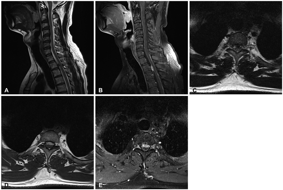
Fig. 1 Preoperative spinal magnetic resonance image. T2-weighted image showed low signal intensity intradural extramedullary mass at T2-3 level (A). Well demarcated enhancing mass in T1-weighted image at T2-3 level was found. The enhancing thick membranous lesion was also found in the dorsal side of the spinal cord from T1 to T5 (B). In the axial view, the mass was located on the ventral side of the spinal cord in T1-weighted (C), T2-weighted (D), and T1-weighted image with gadolinium enhancement (E).
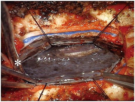
Fig. 2 Intraoperative gross photograph showed a black colored tumor which was diffuse leptomeningeal dissemination through the dorsal surface of the spinal cord. Asterisk (*) indicates cephalad direction.

Fig. 3 Hematoyxlin and eosin stain of the tumor showed melanin pigmentation of tumor cells (A: ×200) and (B: ×400). Positive stain was seen in human melanoma black-45 anti-melanoma monoclonal antibody at mid-portion of the upper portion of figure (C: ×400).
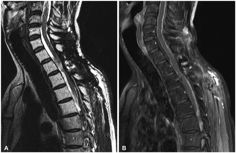
Fig. 4 Spinal magnetic resonance image scan at 22 months after operation. A: Swelling of the spinal cord was seen at the tumor resection site. B: There was no definite tumor mass but diffuse enhancement of leptomenings.
Reference
-
1. Rubin KM. Management of primary cutaneous and metastatic melanoma. Semin Oncol Nurs. 2013; 29:195–205.
Article2. Kim MS, Yoon do H, Shin DA. Primary spinal cord melanoma. J Korean Neurosurg Soc. 2010; 48:157–161.
Article3. Larson TC 3rd, Houser OW, Onofrio BM, Piepgras DG. Primary spinal melanoma. J Neurosurg. 1987; 66:47–49.
Article4. Ryu DS, Park YM, Kim KH, Lee S, Kim KS. Primary intradural extramedullary malignant melanoma in the thoracic spine: case report and literature review. Korean J Spine. 2010; 7:184–187.5. Farrokh D, Fransen P, Faverly D. MR findings of a primary intramedullary malignant melanoma: case report and literature review. AJNR Am J Neuroradiol. 2001; 22:1864–1866.6. Lee CH, Moon KY, Chung CK, et al. Primary intradural extramedullary melanoma of the cervical spinal cord: case report. Spine (Phila Pa 1976). 2010; 35:E303–E307.7. Straathof CS, de Bruin HG, Dippel DW, Vecht CJ. The diagnostic accuracy of magnetic resonance imaging and cerebrospinal fluid cytology in leptomeningeal metastasis. J Neurol. 1999; 246:810–814.
Article8. Chu WC, Lee V, Chan YL, et al. Neurocutaneous melanomatosis with a rapidly deteriorating course. AJNR Am J Neuroradiol. 2003; 24:287–290.9. Davies MA, Liu P, McIntyre S, et al. Prognostic factors for survival in melanoma patients with brain metastases. Cancer. 2011; 117:1687–1696.
Article10. Nishihara M, Sasayama T, Kondoh T, Tanaka K, Kohmura E, Kudo H. Long-term survival after surgical resection of primary spinal malignant melanoma. Neurol Med Chir (Tokyo). 2009; 49:546–548.
Article11. Bidziński J, Kroh H, Leszczyk C, Bojarski P. Primary intraspinal cervical melanoma. Acta Neurochir (Wien). 2000; 142:1069–1070.
Article12. Mirone G, Cinalli G, Spennato P, Ruggiero C, Aliberti F. Hydrocephalus and spinal cord tumors: a review. Childs Nerv Syst. 2011; 27:1741–1749.
Article
- Full Text Links
-
- Actions
-
Cited
- CITED
-
- Close
- Share
-
- Similar articles
-
- A Case of Primary Malignant Leptomeningeal Melanoma of the Spinal Cord: Case Report
- Spinal Cord Malignant Melanoma with Leptomeningeal Metastasis: A Case Report
- Primary Spinal Meningeal Melanocytoma
- An Uncommon Intramedullary Tumor: Primary Spinal Cord Melanoma
- Primary Intradural Extramedullary Malignant Melanoma in the Thoracic Spine: Case Report and Literature Review
