Pituitary Apoplexy Mimicking Meningitis
- Affiliations
-
- 1Department of Neurosurgery, Ajou University School of Medicine, Suwon, Korea. nsksh@ajou.ac.kr
- 2Department of Pathology, Ajou University School of Medicine, Suwon, Korea.
- 3Department of Radiology, Ajou University School of Medicine, Suwon, Korea.
- 4Department of Neurosurgery, Daewoo General Hospital, Geoje, Korea.
- KMID: 2048486
- DOI: http://doi.org/10.14791/btrt.2013.1.2.111
Abstract
- Pituitary apoplexy is a rare but life-threatening disorder. Clinical presentation of this condition includes severe headaches, impaired consciousness, fever, visual disturbance, and variable ocular paresis. The clinical presentation of meningeal irritation is very rare. Nonetheless, if present and associated with fever, pituitary apoplexy may be misdiagnosed as a meningitis. We experienced a case of pituitary apoplexy masquerading as a meningitis. A 42-year-old man presented with meningitis associated symptoms and initial imaging studies did not show evidence of intra-lesional hemorrhage in the pituitary mass. However, a follow-up imaging after neurological deterioration revealed pituitary apoplexy. Hereby, we report our case with a review of literatures.
Keyword
MeSH Terms
Figure
-
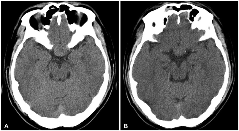
Fig. 1 Non-contrast brain computed tomography scans show a 2.0×1.5 cm sized isodense sellar mass (A), without evidence of intralesional or subarachnoid hemorrhage (B).
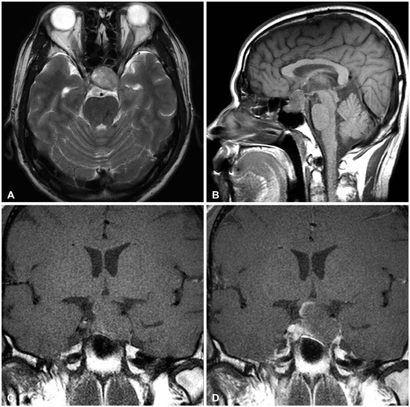
Fig. 2 Initial sellar magnetic resonance imaging findings. T2-weighted axial image shows a sellar mass with heterogenous internal high intensity signals (A). Non-contrast T1-weighted sagittal and coronal images show a pituitary mass extending suprasellar area without typical findings compatible with intralesional hemorrhage (B and C) and contrast enhanced coronal image show a pituitary mass with peripheral enhancement (D).
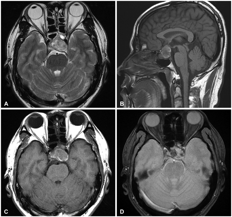
Fig. 3 Follow-up brain magnetic resonance imaging findings. T2-weighted axial image shows mixed signal intensity of pituitary mass (A). Non-contrast T1-weighted sagittal and axial images show peripheral high signal intensity of pituitary mass (B and C). Gradient-echo T2-weighted axial image shows multiple low signal intensities suggesting intralesional hemorrhage (D).

Fig. 4 Histopathologic findings from specimens of the pituitary tumor. Hematoxylin and eosin staining is demonstrating the hemorrhagic necrosis of pituitary gland consistent with pituitary apoplexy (A: ×10 magnification) and pituitary adenoma showing necrotic degeneration with hemorrhage (B: ×200 magnification).
Reference
-
1. Tsitsopoulos P, Andrew J, Harrison MJ. Pituitary apoplexy and haemorrhage into adenomas. Postgrad Med J. 1986; 62:623–626.
Article2. Verrees M, Arafah BM, Selman WR. Pituitary tumor apoplexy: characteristics, treatment, and outcomes. Neurosurg Focus. 2004; 16:E6.
Article3. Nawar RN, AbdelMannan D, Selman WR, Arafah BM. Pituitary tumor apoplexy: a review. J Intensive Care Med. 2008; 23:75–90.4. Brougham M, Heusner AP, Adams RD. Acute degenerative changes in adenomas of the pituitary body--with special reference to pituitary apoplexy. J Neurosurg. 1950; 7:421–439.
Article5. Randeva HS, Schoebel J, Byrne J, Esiri M, Adams CB, Wass JA. Classical pituitary apoplexy: clinical features, management and outcome. Clin Endocrinol (Oxf). 1999; 51:181–188.
Article6. Laws ER. Pituitary tumor apoplexy: a review. J Intensive Care Med. 2008; 23:146–147.
Article7. Cagnin A, Marcante A, Orvieto E, Manara R. Pituitary tumor apoplexy presenting as infective meningoencephalitis. Neurol Sci. 2012; 33:147–149.
Article8. Huang WY, Chien YY, Wu CL, Weng WC, Peng TI, Chen HC. Pituitary adenoma apoplexy with initial presentation mimicking bacterial meningoencephalitis: a case report. Am J Emerg Med. 2009; 27:517.e1–517.e4.
Article9. Bjerre P, Lindholm J. Pituitary apoplexy with sterile meningitis. Acta Neurol Scand. 1986; 74:304–307.
Article10. Dulipsingh L, Lassman MN. Images in clinical medicinePituitary apoplexy. N Engl J Med. 2000; 342:550.
