Bony fusion of the maxilla and mandible as a sequelae of noma: A rare case report
- Affiliations
-
- 1Department of Oral Medicine and Radiology, ITS Center for Dental Studies and Research, Ghaziabad, Uttar Pradesh, India. drshivanandb@yahoo.com
- KMID: 2045028
- DOI: http://doi.org/10.5624/isd.2015.45.3.193
Abstract
- Noma is a gangrenous disease of the orofacial region that leads to severe facial tissue destruction and is a significant cause of death among children. With the advent of modern antibiotics and improved nutrition, children with noma may survive into adulthood, but must face the challenge of undergoing repair of the sequelae of noma. This report describes a case of bony fusion of the maxilla and mandible in a 28-year-old female patient, which was a sequelae of a childhood case of noma.
Keyword
MeSH Terms
Figure
-
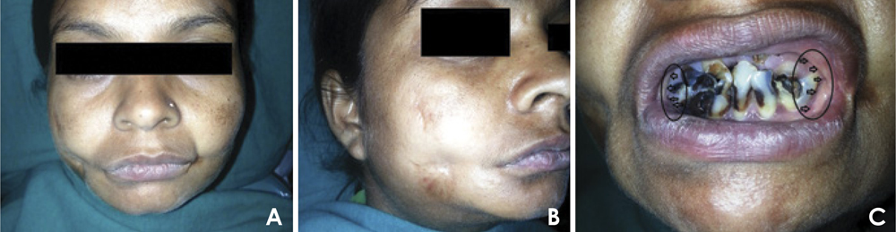
Fig. 1 A. A frontal extraoral photograph shows the scars on both right and left cheeks. B. An extraoral photograph on the right side reveals the scar. C. An intraoral photograph shows grossly decayed anterior teeth with no mouth opening.
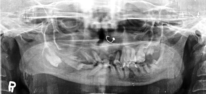
Fig. 2 A panoramic radiograph reveals the fusion of the maxilla and mandible extending from the right upper second premolar to the tip of coronoid process of mandible.
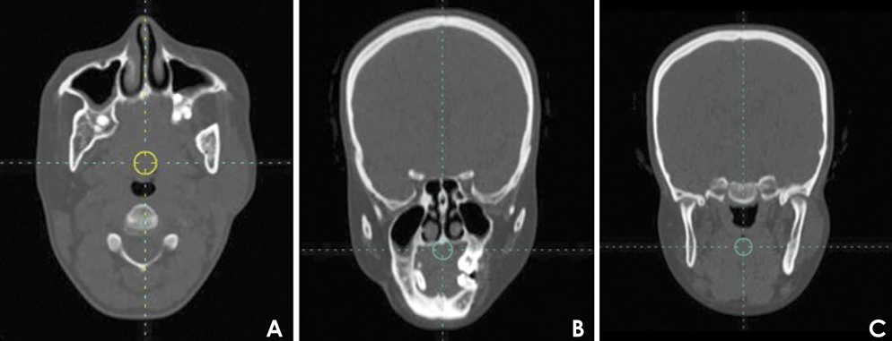
Fig. 3 A. An axial CT image shows the continuous fusion between the maxilla and mandible on the right side. B. A coronal CT image shows the similar features of axial section. C. A coronal CT image shows no fusion of the condylar head of both sides.
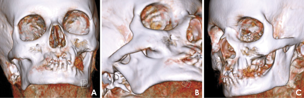
Fig. 4 Three-dimensional CT images. A. Anterior facial area reveals the fused bone. B. Right oblique anterior view indicates bony fusion extending from the right premolar region to the anterior border of ramus of mandible. C. Left oblique anterior view shows no bony fusion.
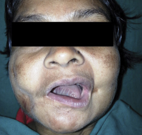
Fig. 5 Post-operative follow-up after 3 month reveals 25 mm mouth opening.
Reference
-
1. Ogbureke KU, Ogbureke EI. NOMA: a preventable "Scourge" of African children. Open Dent J. 2010; 4:201–206.
Article2. Woon CY, Sng KW, Tan BK, Lee ST. Case report journey of a noma face. Eplasty. 2010; 10:e49.3. Baratti-Mayer D, Gayet-Ageron A, Hugonnet S, François P, Pittet-Cuenod B, Huyghe A, et al. Risk factors for noma disease: a 6-year, prospective, matched case-control study in Niger. Lancet Glob Health. 2013; 1:e87–e96.
Article4. Marck KW, Bruijn HP. Surgical treatment of noma. Oral Dis. 1999; 5:167–171.
Article5. Bello SA, Aluko Olokun B, Olaitan AA, Ajike SO. Aetiology and presentation of ankylosis of the temporomandibular joint: report of 23 cases from Abuja, Nigeria. Br J Oral Maxillofac Surg. 2012; 50:80–84.
Article6. Deeb GR, Yih WY, Merrill RG, Lundeen RC. Noma: report of a case resulting in bony ankylosis of the maxilla and mandible. Dentomaxillofac Radiol. 1999; 28:378–382.
Article7. Oluwasanmi JO, Lagundoye SB, Akinyemi OO. Ankylosis of the mandible from cancrum oris. Plast Reconstr Surg. 1976; 57:342–350.
Article8. Marck KW. Noma: a neglected enigma. Lancet Glob Health. 2013; 1:e58–e59.
Article9. Oginni FO, Oginni AO, Ugboko VI, Otuyemi OD. A survey of cases of cancrum oris seen in Ile-Ife, Nigeria. Int J Paediatr Dent. 1999; 9:75–80.
Article10. Wazir SM, Khan SU. Cancrum oris. J Pak Assoc Dermatol. 2008; 18:110–112.11. Lagundoye SB, Oluwasanmi JO. Radiologic examination of trismus as a complication of cancrum oris. Oral Surg Oral Med Oral Pathol. 1975; 39:812–820.
Article12. Lazarus D, Hudson DA. Cancrum oris - a 35-year retrospective study. S Afr Med J. 1997; 87:1379–1382.13. Adekeye EO. Ankylosis of the mandible: analysis of 76 cases. J Oral Maxillofac Surg. 1983; 41:442–449.
Article14. Enwonwu CO, Falkler WA Jr, Phillips RS. Noma (cancrum oris). Lancet. 2006; 368:147–156.
Article15. Giessler GA, Schmidt AB, Deubel U, Cornelius CP. Free flap transfer for closure and interposition-arthroplasty in noma defects of the lateral face associated with bony ankylosis. J Craniofac Surg. 2004; 15:766–773.
Article
