J Rhinol.
2017 May;24(1):48-51. 10.18787/jr.2017.24.1.48.
A Case of Fibromyxoma of the Maxilla
- Affiliations
-
- 1Department of Otorhinolaryngology-Head & Neck Surgery, Dankook University College of Medicine, Cheonan, Korea. entdocjung@hanmail.net
- KMID: 2379790
- DOI: http://doi.org/10.18787/jr.2017.24.1.48
Abstract
- Fibromyxoma is a rare mesenchymal tumor that is benign, but locally invasive. It is a slow-glowing painless tumor with the potential for extensile bony destruction and cortical expansion and shows a relatively high recurrence rate. Fibromyxoma is found predominantly in the jaw, with the mandible more frequently affected than the maxilla. We recently experienced a case of fibromyxoma originating from the maxilla in a 50-year-old woman who complained of swelling on the right side of her cheek. En bloc resection via a sublabial approach and middle meatal antrostomy were performed. A diagnosis of fibromyxoma was based on pathologic findings. No recurrence or locally residual lesion has been found during 2-years follow up. Therefore, we present this rare case with a review of the literature.
Keyword
MeSH Terms
Figure
-
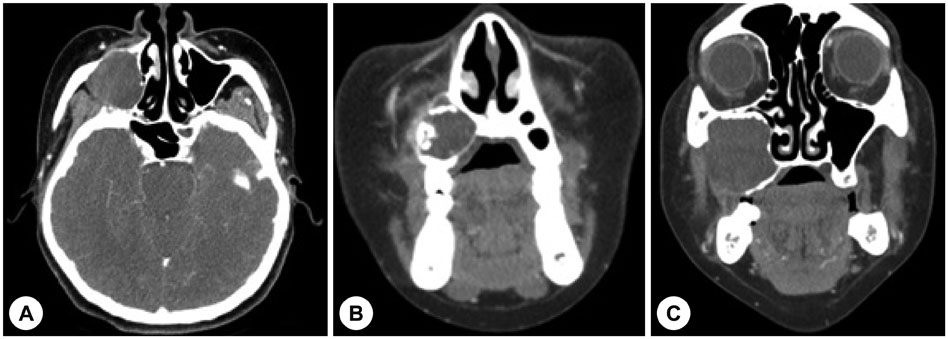
Fig. 1 Axial (A) and coronal (B and C) CT scans show that 3.8×3.5×3.3 cm non-enhancing low attenuated expansile cystic mass with bone erosion nature in the right maxillary sinus.
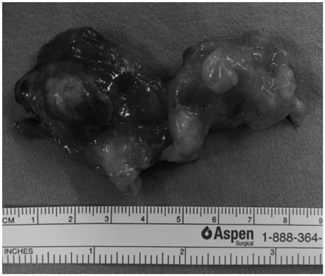
Fig. 2 Gross finding (cutting view in the middle) reveals a grayish gelatinous mass having a glistening mucoid appearance. The mass appears well circumscribed and encapsulated.
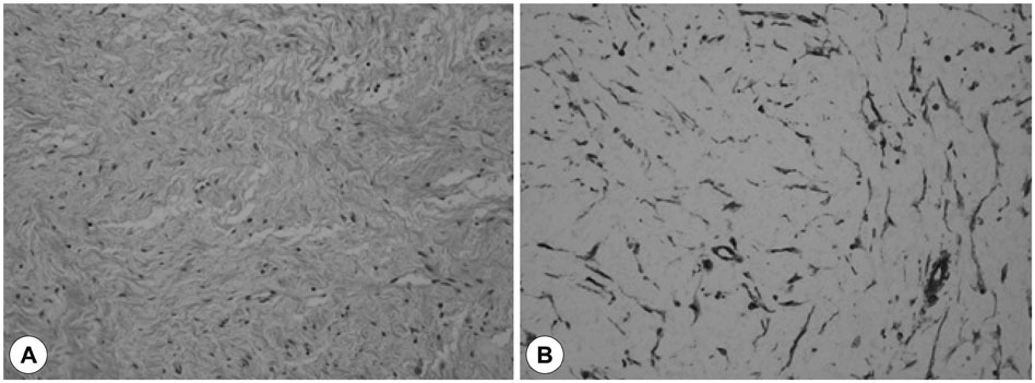
Fig. 3 Histological appearance of fibromyxoma shows that cellular myxoid lesion with bland stellate and spindle cell (A, H & E stain, ×200) and fibromyxoma cells are immunohistochemically positive for vimentin (B).
Reference
-
1. McClure DK, Dahlin DC. Myxoma of bone. Report of three cases. Mayo Clin Proc. 1977; 52(4):249–253.2. Andrews T, Kountakis SE, Maillard AA. Myxomas of the head and neck. Am J Otolaryngol. 2000; 21(3):184–189.
Article3. Chiodo AA, Strumas N, Gilbert RW, Birt BD. Management of odontogenic myxoma of the maxilla. Otolaryngol Head Neck Surg. 1997; 117(6):S73–S76.
Article4. Chuchurru JA, Luberti R, Cornicelli JC, Dominguez FV. Myxoma of the mandible with unusual radiographic appearance. J Oral Maxillofac Surg. 1985; 43(12):987–990.
Article5. Kim YH, Choi JS, Kim BH, Kang SH, Lim DJ, Yu MS. A Large Dentigerous Cyst Found in the Mandible. J Rhinol. 2013; 20(1):46–49.6. Stout AP. Myxoma, the tumor of primitive mesenchyme. Ann Surg. 1948; 127(4):706–719.7. White DK, Chen S, Mohnac AM, Miller AS. Odontogenic myxoma. A clinical and ultrastructural study. Oral Surg Oral Med Oral Pathol. 1975; 39(6):901–917.8. Harris RJ, Garrow E, Spinnato G. Myxoma of the maxilla: report of case. J Oral Surg. 1977; 35(1):70–73.9. Suh SH, Kim SR, Jung YS, Kang HJ. Fibromyxoma Arising from the Maxilla: A Case Report. Korean J Otolaryngol-Head Neck Surg. 1999; 42(10):3121–3125.10. Kim YM, Song SY, Lee CJ. A case of myxoma of the maxillary sinus. Korean J Otolaryngol-Head Neck Surg. 1998; 41(12):1614–1616.11. Kim DH, Jung H, Han TH, Park SK. Myxoma of the Maxillary Sinus: A Case Report. Korean J Otolaryngol-Head Neck Surg. 2007; 50(1):76–79.12. Park J, Dhong HJ, Chung SK, Kim HY. Myxoma of Sphenoid Sinus. J Rhinol. 2012; 19(2):141–144.13. Pahl S, Henn W, Binger T, Stein U, Remberger K. Malignant odontogenic myxoma of the maxilla: case with cytogenetic confirmation. J Laryngol Otol. 2000; 114(7):533–535.
Article
