Osteoblastoma-like Osteosarcoma of the Proximal Tibia: A Case Report
- Affiliations
-
- 1Department of Radiology, Myoungji Hospital, Kwandong University College of Medicine, Korea.
- 2Department of Radiology, Kangbuk Samsung Hospital, Sungkyunkwan University School of Medicine, Korea. parkhiji@gmail.com
- 3Department of Pathology, Myoungji Hospital, Kwandong University College of Medicine, Korea.
- 4Department of Orthpedic Surgery, Myoungji Hospital, Kwandong University College of Medicine, Korea.
- KMID: 2040819
- DOI: http://doi.org/10.3348/jksr.2011.64.5.497
Abstract
- Osteoblastoma-like osteosarcoma is a rare, low-grade variant of osteosarcoma that may resemble osteoblastoma clinically, radiographically and histologically. We report a case of osteoblastoma-like osteosarcoma in the right proximal tibia in a 17-year-old woman. Plain radiography revealed an irregular osteolytic lesion in the metaepiphysis of the right proximal tibia and transverse fracture line of the metaphysic through an osteolytic lesion. MRI showed well-defined mass confined to the medullary space with cortical disruption and pathologic fracture. The MR signals were low on T1-weighted image and intermediate on T2-weighted image.
MeSH Terms
Figure
-
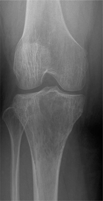
Fig. 1 Initial anteroposterior radiograph reveals an irregular osteolytic lesion in the metaepiohysis of the proximal tibia.
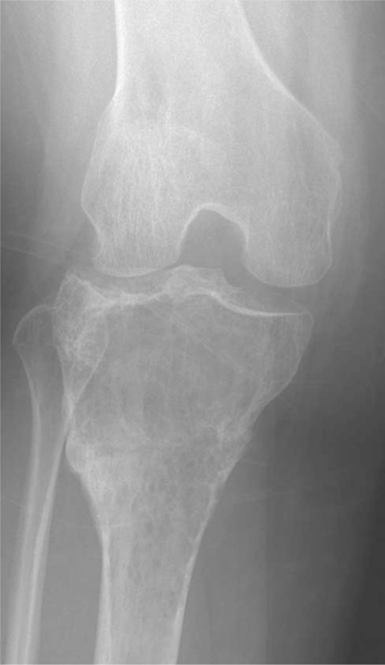
Fig. 2 A year later, tumor lesion aggravates and pathologic fracture is combined.

Fig. 3 A, B. Coronal T1 (TR 590, TE 15) and T2 (TR 4011, TE 100)-weighted MR image shows low and intermediate signal intensity lesion confined to the tibia. C. Fat-suppressed axial T2-weighted image (TR 3200, TE 60) predominatly showing hyperintense lesion with clear circumscription and no prominent periosteal reaction.
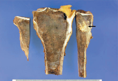
Fig. 4 Photograph of the gross specimen shows the tumor, measuring 13 cm in length, in the tibia. The cut surfaces reveal relatively well circumscribed lesion abutting the articular cartilage. Grafted bone cement is present (arrow).
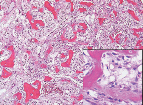
Fig. 5 Microphotograph of the tumor. The tumor is composed of woven bone, trabeculae lined by plump osteoblastic tumor cells, and a stroma rich in vessels (×100, inset: original magnification ×600).
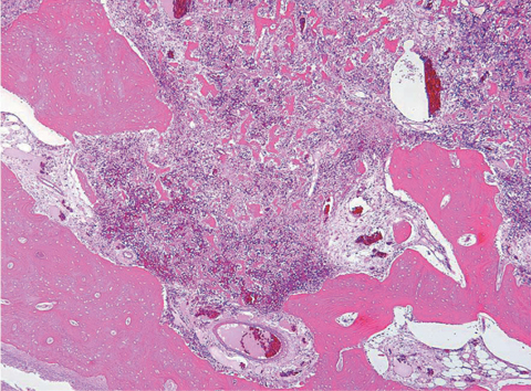
Fig. 6 Microphotograph of the tumor. The tumor tissue permeation through the host bony trabeculae is evident in the periphery (×40).
Reference
-
1. Abramovici L, Kenan S, Hytiroglou P, Rafii M, Steiner GC. Osteoblastoma-like osteosarcoma of the distal tibia. Skeletal Radiol. 2002; 31:179–182.2. Bonar SF, McCarthy S, Stalley P, Schatz J, Soper J, Scoyler R, et al. Epiphyseal osteoblastoma-like osteosarcoma. Skeletal Radiol. 2004; 33:46–50.3. Tani T, Okada K, Shoji K, Hashimoto M, Sageshima M. Osteoblastoma-like osteosarcoma. Skeletal Radiol. 2000; 29:656–659.4. Kumar NL, Rosenberg AE, Raskin KA. Osteoblastoma-like osteosarcoma of the cuboid: a case report. J Orthop Surg Res. 2010; 5:52.5. Bertoni F, Unni KK, Mcleod RA, Dahlin DC. Osteosarcoma resembling osteoblastoma. Cancer. 1985; 55:416–426.6. Bertoni F, Bacchini P, Donati D, Martini A, Picci P, Campanacci M. Osteoblastoma-like osteosarcoma. The Rizzoli Institute experience. Mod Pathol. 1993; 6:707–716.7. Dorfman HD, Weiss S. Borderline osteoblastic tumors: problems in differential diagnosis of aggressive osteoblastoma and low grade osteosarcoma. Semin Diagn Pathol. 1984; 1:215–234.
